[Preprint]. 2025 Jun 1:2025.05.31.654582.
doi: 10.1101/2025.05.31.654582.

Integrin-Specific Signaling Drives ER Stress-Dependent Atherogenic Endothelial Activation

Affiliations

Integrin-Specific Signaling Drives ER Stress-Dependent Atherogenic Endothelial Activation

Cyrine Ben Dhaou et al. bioRxiv. .

Abstract

Atherogenic endothelial activation arises from both the local arterial microenvironment-characterized by altered extracellular matrix composition and disturbed blood flow-and soluble proinflammatory stimuli such as oxidized low-density lipoprotein (oxLDL). Fibronectin, a provisional extracellular matrix protein enriched at atheroprone sites, enhances endothelial activation and inflammation triggered by oxLDL and disturbed flow. Although endoplasmic reticulum (ER) stress contributes to vascular dysfunction, the role of matrix composition in regulating ER stress remains unknown. We show that oxLDL and disturbed flow induce ER stress selectively in endothelial cells adhered to fibronectin, whereas both stimuli fail to induce ER stress in cells on basement membrane proteins. This matrix-specific ER stress response requires integrin activation, as endothelial cells deficient for integrin activation (talin1 L325R mutation) fail to activate ER stress in response to disturbed flow and oxLDL and direct stimulation of integrin activation using CHAMP peptides is sufficient to trigger ER stress. Blunting endothelial expression of fibronectin-binding integrins (α5, αv) using siRNA prevents ER stress in response to atherogenic stimuli in vitro, whereas endothelial α5 and αv deletion reduces ER stress at atheroprone sites in vivo. The mechanisms driving integrin-dependent ER stress remain unclear, since matrix composition does not affect protein translation, unfolded protein accumulation, or superoxide production, and scavenging superoxide (TEMPOL) does not reduce integrin-dependent ER stress. Inhibiting ER stress with TUDCA reduces proinflammatory and metabolic gene expression (bulk RNAseq) but does not prevent NF-κB activation, a classic proinflammatory transcription factor. Rather, TUDCA prevents activation of c-jun N-terminal kinase (JNK) and c-jun activation, and blocking JNK (SP600126) or c-Jun activity (TAM67) prevents proinflammatory gene expression following both stimuli. Together, these findings offer new insight into how the arterial microenvironment contributes to atherogenesis, with fibronectin-binding integrin signaling promotes ER stress in response to mechanical and metabolic stressors, thereby amplying proinflammatory endothelial activation through JNK-c-Jun signaling.

Keywords: Atherosclerosis; ER Stress; Inflammation; Integrin; Oxidized LDL; Shear Stress.


Conflict of interest statement

Disclosures The authors declare that the research was conducted in the absence of any commercial or financial relationships that could be construed as a potential conflict of interest.

Figures

Figure 1:
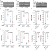
Figure 1:. ER stress exhibits matrix specificity in response to OxLDL and disturbed flow.
(A-F) Human aortic endothelial cells (HAECs) plated on fibronectin or basement membrane were treated with OxLDL (100 μg, 24 h) or (G-L) subjected to oscillatory shear stress (OSS; ±5 dynes/cm2 with 1 dyne/cm2 forward flow, 18h). ER stress markers (XBP1s, P-eIF2α, ATF4, BIP and NRF2) were assessed by Western blotting and normalized to total protein levels using GAPDH as the loading control. (M) HAECs plated on fibronectin or basement membrane were treated with OxLDL (100 μg, 24 h) or (N) subjected to OSS (±5 dynes/cm2 with 1 dyne/cm2 forward flow, 18h), and stained for XBP1s. Scale bar: 20 μm. (O) HAECs plated on fibronectin or basement membrane were transfected with XBP1s-mNeoGreen, or (P) with ATF4-mScarlet constructs, and treated with OxLDL (100 μg) for the indicated times. Scale bar: 20 μm. (Q) HAECs plated on fibronectin or basement membrane were transfected with either XBP1s-mNeoGreen, or (R) ATF4-mScarlet constructs, and subjected to OSS (±5 dynes/cm2 with 1 dyne/cm2 forward flow, 18h). Scale bar: 20 μm. P values were determined by two-way ANOVA with Tukey’s multiple comparisons test. Data are presented as mean ± SEM. Each point represents one independent experiment.
Figure 2:
Figure 2:. Integrin activation drives ER stress.
(A-C) Murine lung endothelial cells (MLECs) were isolated from Talin WT and Talin1 L325R, plated on fibronectin, and treated with OxLDL (100 μg, 24 h) or (D-F) subjected to OSS (±5 dynes/cm2 with 1 dyne/cm2 forward flow; 18h). ER stress markers (XBP1s and P-eIF2α) were assessed by Western blotting and normalized to total protein levels using GAPDH as the loading control. (G-K) HAECs were treated with either controls or activating α5β1 or αvβ3 CHAMPs (4uM, 24 h). ER stress markers (XBP1s and P-eIF2α) were assessed by Western blotting and normalized to total protein levels using GAPDH as the loading control. P values were determined by two-way ANOVA with Tukey’s multiple comparisons test for B-F and a nonparametric Mann-Whitney test for H-K. Data are presented as mean ± SEM. Each point represents one independent experiment.
Figure 3:
Figure 3:. Differential role of fibronectin-binding integrins in regulating endothelial ER stress.
(A-C) HAECs were transfected with siRNA targeting α5 or αv and subjected to OSS (±5 dynes/cm2 with 1 dyne/cm2 forward flow, 18h). ER stress markers (XBP1s and P-eIF2α) were assessed by Western blotting and normalized to total protein levels using GAPDH as the loading control. (D-F) HAECs were transfected with siRNA targeting β3 subunit and treated with OxLDL (100 μg, 24 h). ER stress markers (XBP1s and P-eIF2α) were assessed by Western blotting and normalized to total protein levels using GAPDH as the loading control. (G-I) HAECs were transfected with siRNA targeting α5 and treated with OxLDL (100 μg, 24 h). ER stress markers (XBP1s and P-eIF2α) were assessed by Western blotting and normalized to total protein levels using GAPDH as the loading control. P values were determined by two-way ANOVA with Tukey’s multiple comparisons test. Data are presented as mean ± SEM. Each point represents one independent experiment.
Figure 4:
Figure 4:. Deletion of fibronectin-binding integrins in endothelium blocks ER stress in mice.
(A) Representative images of aortic arches from α5 and αv knockout (KO) mice and their wild-type controls, fed a high-fat diet (HFD) for 2 weeks, stained for XBP1s and CD31. Scale bars: 20 μm. (B) Quantification of the percentage of endothelium positive for XBP1s using NIS Elements software (n=4). (C) Representative images of brachiocephalic arteries from α5 and αv KO mice and their wild-type controls, fed an HFD for 8 weeks, stained for XBP1s and CD31. Scale bars: 20 μm. (D) Quantification of the percentage of endothelium positive for XBP1s using NIS Elements software (n=3–6). P values were determined by one-way ANOVA with Dunnett’s multiple comparisons test. Data are presented as violin plots, with the shaded area representing the 5th to 95th percentile and the central dot indicating the median. Each point represents one animal.
Figure 5:
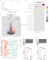
Figure 5:. Blocking ER stress inhibits fibronectin-specific OxLDL and disturbed flow-mediated endothelial inflammation.
(A) Principal component analysis (PCA) of HAECs plated on fibronection, pretreated with TUDCA (20uM, 2h) and exposed to OSS (±5 dynes/cm2 with 1 dyne/cm2 forward flow, 18h). (B) Volcano plot of differentially expressed genes (DEGs) in HAECs pretreated with TUDCA (20uM, 2h) and exposed to OSS compared to untreated HAECs exposed to OSS (18 h). (C) Bubble plot of KEGG pathway enrichment analysis of downregulated DEGs in HAECs pretreated with TUDCA (20uM, 2h) and exposed to OSS compared to untreated HAECs exposed to OSS (±5 dynes/cm2 with 1 dyne/cm2 forward flow, 18h). (D) Heatmap of KEGG pathways comparing RNA sequencing of HAECs pretreated with TUDCA and exposed to OSS compared to untreated HAECs exposed to OSS (±5 dynes/cm2 with 1 dyne/cm2 forward flow, 18h). (E-F) HAECs plated on fibronectin were pretreated with TUDCA (20uM, 2h), treated with OxLDL (100 μg, 24 h) or (G-H) subjected to OSS (±5 dynes/cm2 with 1 dyne/cm2 forward flow, 18h). ICAM-1 and VCAM-1 were assessed by Western blotting and normalized to total protein levels using GAPDH as the loading control. P values were determined by two-way ANOVA with Tukey’s multiple comparisons test. Data are presented as mean ± SEM. Each point represents one independent experiment.
Figure 6:
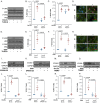
Figure 6:. ER stress promotes endothelial activation via JNK signaling.
(A-C) HAECs plated on fibronectin were pretreated with TUDCA (20uM, 2h), treated with OxLDL (100 μg, 24 h) or (D-F) subjected to OSS (±5 dynes/cm2 with 1 dyne/cm2 forward flow, 18h). P-JNK and P-P65 were assessed by Western blotting and normalized to total protein levels using GAPDH as the loading control. (G-H) HAECs plated on fibronectin were pretreated with SP600125 (1uM, 2h), treated with OxLDL (100 μg, 24 h) or (I-J) subjected to OSS (±5 dynes/cm2 with 1 dyne/cm2 forward flow, 18h). VCAM-1 was assessed by Western blotting and normalized to total protein levels using GAPDH as the loading control. (K) HAECs plated on fibronectin were pretreated with TUDCA, exposed to OxLDL (100 μg, 24 h) or (I) subjected to OSS (±5 dynes/cm2 with 1 dyne/cm2 forward flow), and stained for p-c-JUN and phalloidin. Scale bar: 20 μm. (M-N) HAECs plated on fibronectin transfected with TAM67, treated with OxLDL (100 μg, 24 h) or (O-P) subjected to OSS (±5 dynes/cm2 with 1 dyne/cm2 forward flow, 18h). VCAM-1 was assessed by Western blotting and normalized to total protein levels using GAPDH as the loading control. P values were determined by two-way ANOVA with Tukey’s multiple comparisons test. Data are presented as mean ± SEM. Each point represents one independent experiment.

References

    1. Martin S.S., Aday A.W., Allen N.B., Almarzooq Z.I., Anderson C.A.M., Arora P., Avery C.L., Baker-Smith C.M., Bansal N., Beaton A.Z., Commodore-Mensah Y., Currie M.E., Elkind M.S.V., Fan W., Generoso G., Gibbs B.B., Heard D.G., Hiremath S., Johansen M.C., Kazi D.S., Ko D., Leppert M.H., Magnani J.W., Michos E.D., Mussolino M.E., Parikh N.I., Perman S.M., Rezk-Hanna M., Roth G.A., Shah N.S., Springer M.V., St-Onge M.-P., Thacker E.L., Urbut S.M., Van Spall H.G.C., Voeks J.H., Whelton S.P., Wong N.D., Wong S.S., Yaffe K., Palaniappan L.P., American Heart Association Council on Epidemiology and Prevention Statistics Committee and Stroke Statistics Committee, 2025 Heart Disease and Stroke Statistics: A Report of US and Global Data From the American Heart Association, Circulation (2025). 10.1161/CIR.0000000000001303. - DOI
    1. GBD 2019 Diseases and Injuries Collaborators, Global burden of 369 diseases and injuries in 204 countries and territories, 1990–2019: a systematic analysis for the Global Burden of Disease Study 2019, Lancet 396 (2020) 1204–1222. 10.1016/S0140-6736(20)30925-9. - DOI - PMC - PubMed
    1. Ahmad F.B., Cisewski J.A., Anderson R.N., Mortality in the United States - Provisional Data, 2023, MMWR Morb Mortal Wkly Rep 73 (2024) 677–681. 10.15585/mmwr.mm7331a1. - DOI - PMC - PubMed
    1. Cheng C., Tempel D., van Haperen R., van der Baan A., Grosveld F., Daemen M.J.A.P., Krams R., de Crom R., Atherosclerotic lesion size and vulnerability are determined by patterns of fluid shear stress, Circulation 113 (2006) 2744–2753. 10.1161/CIRCULATIONAHA.105.590018. - DOI - PubMed
    1. Sorescu G.P., Song H., Tressel S.L., Hwang J., Dikalov S., Smith D.A., Boyd N.L., Platt M.O., Lassègue B., Griendling K.K., Jo H., Bone morphogenic protein 4 produced in endothelial cells by oscillatory shear stress induces monocyte adhesion by stimulating reactive oxygen species production from a nox1-based NADPH oxidase, Circ Res 95 (2004) 773–779. 10.1161/01.RES.0000145728.22878.45. - DOI - PubMed

Publication types