[Preprint]. 2025 May 29:2025.05.29.656827.
doi: 10.1101/2025.05.29.656827.

Proximity biotinylation at the host- Shigella interface reveals UFMylation as an antibacterial pathway

Affiliations

Proximity biotinylation at the host- Shigella interface reveals UFMylation as an antibacterial pathway

Ana T López-Jiménez et al. bioRxiv. .

Abstract

Host cells contest invasion by intracellular bacterial pathogens with multiple strategies that recognise and / or damage the bacterial surface. To identify novel host defence factors targeted to intracellular bacteria, we developed a versatile proximity biotinylation approach coupled to quantitative mass spectrometry that maps the host-bacterial interface during infection. Using this method, we discovered that intracellular Shigella and Salmonella become targeted by UFM1-protein ligase 1 (UFL1), an E3 ligase that catalyses the covalent attachment of Ubiquitin-fold modifier 1 (UFM1) to target substrates in a process called UFMylation. We show that Shigella antagonises UFMylation in a dual manner: first, using its lipopolysaccharide (LPS) to shield from UFL1 recruitment; second, preventing UFM1 decoration by the bacterial effector IpaH9.8. Absence of UFMylation leads to an increase of bacterial burden in both human cells and zebrafish larvae, suggesting that UFMylation is a highly conserved antibacterial pathway. Contrary to canonical ubiquitylation, the protective role of UFMylation is independent of autophagy. Altogether, our proximity mapping of the host-bacterial interface identifies UFMylation as an ancient antibacterial pathway and holds great promise to reveal other cell-autonomous immunity mechanisms.

Keywords: E3 ligases; Shigella; UFMylation; bacterial infection; cell-autonomous immunity; proximity biotinylation; ubiquitin-like systems.


Figures
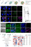
Figure 1.
Figure 1.. Novel proximity biotinylation approach to identify the S. flexneri proxisome during infection.
a, Strategy for the in vitro functionalisation of S. flexneri for proximity mapping. First, an anti-GFP nanobody is displayed on the S. flexneri surface by induction with IPTG. After that, S. flexneri is coated with purified GFP-APEX2 in vitro. Then, functionalised bacteria are used for infection assays. Finally, the addition of biotin phenol (BP) and H2O2 enables the proximity biotinylation reaction during infection. b, Representative airyscan confocal images showing biotinylation in the vicinity of functionalised S. flexneri during infection in HeLa cells, in the presence or absence of BP and H2O2. SAV stands for streptavidin. Scale bar, 5 μm and 1 μm for the inset. c, Western blot shows a smear of biotinylated proteins in HeLa cells infected with functionalised S. flexneri upon addition of BP and H2O2. d, Proximity biotinylation occurs in zebrafish larvae at 45 minutes post infection of functionalised S. flexneri at the tail musculature. The transgenic zebrafish line Tg(lyzC:DsRed2)nz50 was used to visualise fluorescent neutrophils. Scale bar, 50 μm and 10 μm for the inset. e, Experimental workflow to identify the proxisome of S. flexneri WT and ΔmxiE mutant using mass spectrometry. Image created using BioRender. f, Heatmap of E3 ligases found in the proxisomes of S. flexneri during infection, inversely ordered by p-value.
Figure 2.
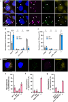
Figure 2.. Several members of the UFMylation pathway are recruited to S. flexneri and S. Typhimurium during infection.
a, Representative confocal images of HeLa cells infected with S. flexneri WT and ΔrfaC. Arrows show UFL1 recruitment to intracellular bacteria. Scale bar, 10 μm. b, Percentage of S. flexneri WT (n=1,311) and ΔrfaC (n=630) colocalising with UFL1 and ubiquitin (FK2), from at least 3 biological replicates. Two-way ANOVA and Tukey’s multiple comparison test. c, Percentage of S. flexneri WT (n=882) and ΔrfaC (n=643) colocalising with UFM1 and ubiquitin (FK2), from at least 3 biological replicates. Two-way ANOVA and Tukey’s multiple comparison test. d, Representative confocal image of HeLa cell infected with S. Typhimurium and stained for UFL1 and ubiquitin (FK2). Scale bar, 10 μm. e, Percentage of S. Typhimurium (n=740) colocalising with UFL1 and ubiquitin (FK2) f, Percentage of S. Typhimurium (n=823) colocalising with UFM1 and ubiquitin (FK2). g, Percentage of S. Typhimurium (n=735) colocalising with UFL1 and GFP-RNF213. The results are represented as mean ± SD.
Figure 3.
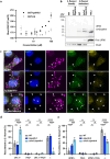
Figure 3.. The S. flexneri secreted effector IpaH9.8 prevents UFM1 conjugation of intracellular bacteria.
a, Protein-protein interaction assay monitoring fluorescence polarisation (FP) of rhodamine-labelled UFM1 in response to increasing concentrations of recombinant GST-IpaH9.8. To control for possible interactions with the GST tag of GST-IpaH9.8, GST-ubiquitin is titrated instead. b, Western blot showing UFM1 conjugates in S. flexneri WT and ΔrfaCΔipaH9.8 purified from infection that are absent in bacteria from broth. UFM1 species present in uninfected HeLa cells are shown as a control. Different exposure times were used to detect free UFM1 (low) and UFM1 conjugates (high) due to relative differences in abundance, using anti-UFM1 antibodies (Abcam). c, Representative confocal images of HeLa cells infected with S. flexneri WT, ΔipaH9.8 and ΔrfaCΔipaH9.8. Arrows show UFL1 recruitment to intracellular bacteria. Scale bar, 10 μm. d, Percentage of S. flexneri WT (n=1,311), ΔipaH9.8 (n=463) and ΔrfaCΔipaH9.8 (n=553) colocalising with UFL1 and ubiquitin (FK2). e, Percentage of S. flexneri WT (n=882), ΔipaH9.8 (n=476) and ΔrfaCΔipaH9.8 (n=526) colocalising with UFM1 and ubiquitin (FK2). Two-way ANOVA and Tukey’s multiple comparison test. UFL1 and UFM1 quantifications for WT S. flexneri are the same as presented in Fig. 2b, c. The results are represented as mean ± SD.
Figure 4.
Figure 4.. UFMylation has a conserved antibacterial role independent of autophagy.
a, Representative confocal microscopy images of HeLa cells infected with S. flexneri mCherry and immunostained against UFL1 and UFM1. Arrows point at intracellular bacteria colocalising with UFL1 and UFM1 with a loss of mCherry fluorescence. Scale bar, 10 μm and 1 μm for the inset. b, Representative microscopy image of HeLa cells expressing GFP-LC3 infected with S. flexneri mCherry and immunostained against UFL1. Arrows indicate intracellular S. flexneri that recruit UFL1 and lose their mCherry fluorescence but are not engulfed in an autophagosome. Scale bar, 10 μm and 1 μm for the inset. c, Quantification of UFL1 and GFP-LC3 recruitment to S. flexneri WT (n=655), ΔipaH9.8 (n=733), ΔrfaC (n=422), and double ΔrfaCΔipaH9.8 (n=627) mutants. Two-way ANOVA and Tukey’s multiple comparison test. d, Quantification of UFL1 and GFP-LC3 recruitment to S. Typhimurium (n=582). e, Intracellular growth of S. flexneri WT, ΔrfaCΔipaH9.8 and S. Typhimurium in HeLa cells treated with ufm1 siRNA normalised to control siRNA after 5 hours post infection. f, Zebrafish larvae survival after S. flexneri infection at the caudal vein for wildtype larvae (46.40%) and ufl1 (21.85%) and ufm1 (18.00%) KD crispants. Data was obtained from 76 wildtype, 81 ufl1 and 70 ufm1 KD crispant zebrafish larvae from two biological replicates. g, S. flexneri recovered from infected zebrafish larvae at 48 hours post infection (h.p.i) for wildtype larvae, ufl1 and ufm1 KD crispants. h, Zebrafish larvae survival after S. Typhimurium infection at the caudal vein for wildtype larvae (63.15%), ufl1 (31.81%) and ufm1 (27.91%) KD crispants. Data was obtained from 54 wildtype, 61 ufl1 and 55 ufm1 KD crispant zebrafish larvae from two biological replicates. i, S. Typhimurium recovered from infected zebrafish larvae at 48 hours post infection, for wildtype larvae, ufl1 and ufm1 KD crispants. The results are represented as mean ± SD.

References

    1. Khalil I. A. et al. Morbidity and mortality due to Shigella and enterotoxigenic Escherichia coli diarrhoea: the Global Burden of Disease Study 1990–2016. Lancet Infect Dis 18, 1229–1240 (2018). - PMC - PubMed
    1. Bajunaid W. et al. The T3SS of Shigella: expression, structure, function, and role in vacuole escape. Microorganisms Vol. 8, Page 1933 8, 1933 (2020). - PMC - PubMed
    1. Pinaud L. et al. Identification of novel substrates of Shigella T3SA through analysis of its virulence plasmid-encoded secretome. PLoS One 12, e0186920 (2017). - PMC - PubMed
    1. Ray K. et al. Tracking the dynamic interplay between bacterial and host factors during pathogen-induced vacuole rupture in real time. Cell Microbiol 12, 545–556 (2010). - PubMed
    1. Bernardini M. L., Mounier J., D’Hauteville H., Coquis-Rondon M. & Sansonetti P. J. Identification of icsA, a plasmid locus of Shigella flexneri that governs bacterial intra- and intercellular spread through interaction with F-actin. Proc Natl Acad Sci U S A 86, 3867–3871 (1989). - PMC - PubMed

Publication types

LinkOut - more resources