[Preprint]. 2025 Jun 2:2025.04.11.648421.
doi: 10.1101/2025.04.11.648421.

A noncanonical Pol III-dependent, Microprocessor-independent biogenesis pathway generates a germline enriched miRNA family

Affiliations

A noncanonical Pol III-dependent, Microprocessor-independent biogenesis pathway generates a germline enriched miRNA family

Rima M Sakhawala et al. bioRxiv. .

Update in

Abstract

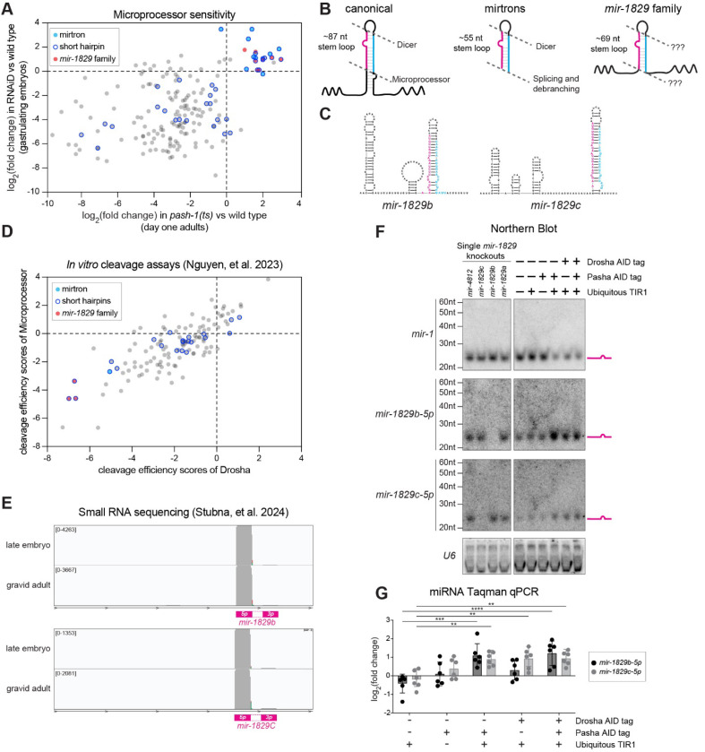
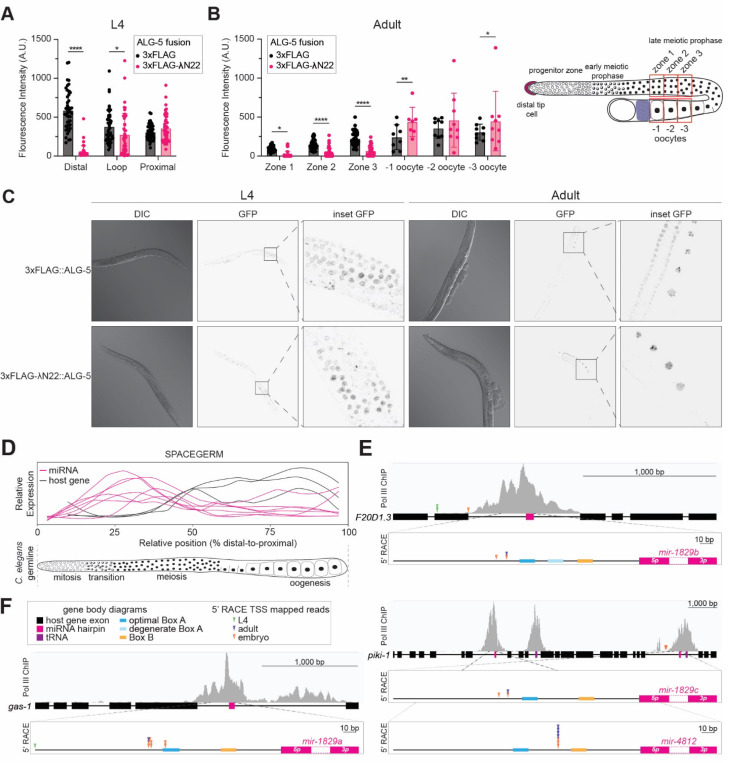
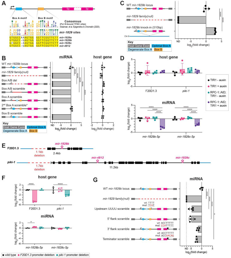
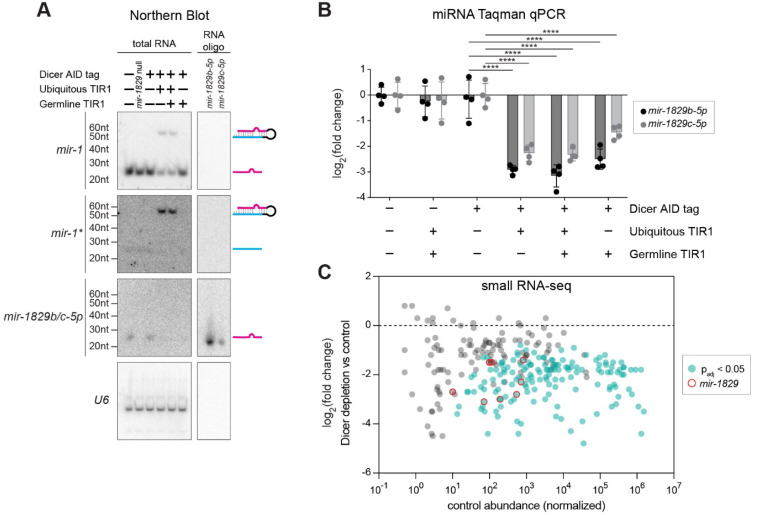
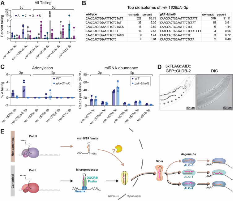
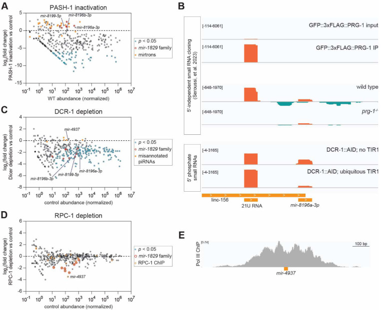
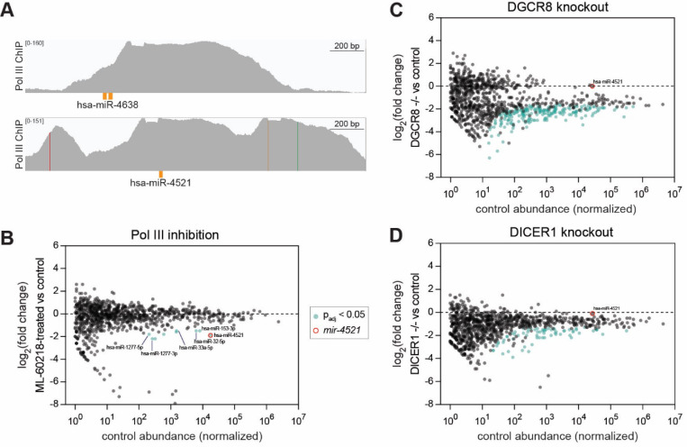
MicroRNAs (miRNAs) are short RNAs that post-transcriptionally regulate gene expression. In canonical miRNA biogenesis, primary miRNAs are transcribed from intergenic loci or intronic regions by RNA polymerase II, sequentially cleaved by the Microprocessor complex and Dicer, and resulting mature miRNAs are loaded into Argonaute to repress target mRNAs. A minority of miRNAs are generated via noncanonical biogenesis pathways that bypass the Microprocessor and/or Dicer. Here, we describe a new Pol III-dependent, Microprocessor-independent, and Dicer-dependent biogenesis pathway exemplified by the mir-1829 family in C. elegans. Although the mir-1829 family loci reside in intronic regions of protein-coding genes, we show that the miRNAs are derived from independent Pol III transcripts. Unlike other Pol III-dependent miRNAs, the mir-1829 family small RNAs are the dominant species derived from their loci, rather than fragments of a larger functional noncoding RNA. These germline-enriched miRNAs are loaded in multiple miRNA Argonautes, including the recently-characterized germline Argonaute ALG-5, which we demonstrate is repressive when tethered to a reporter transcript. We extend these findings, identifying additional Pol III-transcribed and noncanonical small RNAs in C. elegans and human datasets, including human miR-4521. These young, noncanonical miRNAs may represent an early snapshot in the evolution of de novo miRNA genes.

Keywords: DGCR8; Drosha; Pasha; RNA Polymerase III; miR-1277; miR-153; miR-32; miR-33a; mir-8196; pash-1.


Conflict of interest statement

Competing Interest Statement The authors have no competing interests to declare.

Figures

Figure 1.
Figure 1.. The mir-1829 family is Microprocessor-independent.
A) Comparison of log2(fold change) of miRNAs 24h post-upshift to restrictive temperature in pash-1(ts) vs. wild type (Vieux et al. 2021) (x-axis) to those in RNAiD depletion of the Microprocessor (Dexheimer et al. 2020) (y-axis). miRNAs are annotated as having a “short hairpin” if predicted structure on MirGeneDB has <32bp (Fromm et al. 2020). A small number of miRNAs are insensitive to Microprocessor depletion in both datasets, including mirtrons and the mir-1829 family. Only miRNAs with baseMean ≥ 10 in the pash-1(ts) data set are plotted. B) Schematics of miRNA structures emphasizing long basal stem of canonical miRNAs and lack thereof in mirtrons and the mir-1829 family. C) Predicted secondary structures of the pri-mir-1829b and pri-mir-1829c. D) Cleavage efficiency scores of Drosha (x-axis) or Microprocessor (y-axis) was calculated as log2(∑NP + 0.1) – log2(∑NS + 0.1), where NP and NS are normalized counts of cleaved products and pri-miRNA substrates, respectively (Nguyen et al. 2023). The mir-1829 family is among the least favorable substrates. E) Genome browser tracks showing raw sequence reads from bias-minimized small RNA cloning from Stubna, et al. 2024. F) Northern blot of total RNA from adult animals exposed to auxin for 24h starting at the L4 stage at 20°C. mir-1829b-5p, mir-1829c-5p, and U6 probes were 5’ end-labelled, whereas mir-1 probe was labelled according to Starfire method (Behlke et al. 2000). High stringency conditions were used for probing; see methods for details. G) Same samples used for northern blotting were assayed by RT-qPCR. miRNA expression is normalized to a small RNA control (sn2429), then further normalized to wild type samples. Mean and standard deviation of six biological replicates shown. Two-way ANOVA (****p<0.0001, ***p<0.001, **p<0.01).
Figure 2.
Figure 2.. ALG-5-mediated reporter repression and mir-1829 family expression both peak in the distal germline.
A) Measured GFP fluorescence intensity from five nuclei per region (distal, loop, and proximal) across nine L4-staged animals, grown at 20°C expressing a germline single-copy GFP::histone transgene with three boxB RNA hairpins in its 3 UTR. CRISPR-generated animals expressing ALG-5 fusion proteins were tagged with either 3xFLAG and λN22 (experimental) or 3xFLAG alone (control) in a mut-2(null) background. B) Measured relative GFP fluorescence intensity in Day 1 adult worms (same strains as in panel A). Average fluorescence intensity was measured from six regions of interest (shown in right schematic). Zones 1–3 correspond to regions of the distal gonad across from the –1, –2, and –3 oocytes, respectively. Five nuclei were measured in each zone in the distal germline per nine adult worms. A-B) Mean and SD shown. Two-way ANOVA (****p<0.0001, **p<0.01, *p<0.05). C) Representative images of A and B. D) Schematic of relative expression levels of the mir-1829 family and its host genes in the C. elegans germline. Data from SPACEGERM (Diag et al. 2018) indicates that the mir-1829 family (pink) peaks in expression in the distal germline, whereas its host genes (black) peak in expression in the proximal germline. E-F) Prominent Pol III ChIP-seq peaks (Araya et al. 2014) reside at the mir-1829 loci (pink). Arrowheads: 5’ RACE reads cloned from RNA of indicated stage. Putative Pol III promoter sequence motifs upstream of the miRNA precursors are indicated.
Figure 3.
Figure 3.. mir-1829 family members are transcribed by Pol III.
A) MUSCLE alignment of the mir-1829 family putative Pol III promoter sequence motifs. B) Schematic of CRISPR alleles generated at the mir-1829b locus (left) and qPCR of resulting miRNA and host gene expression (right) from young adult samples in wildtype background, grown at 25°C. C) Schematic of genomic sources of mir-1829b (left), including re-integration of a 510bp minimal mir-1829b transcriptional unit in a mir-1829 family(null) mutant background (bottom left). qPCR of mir-1829b expression from young adult samples grown at 25°C (right). D) qPCR of miRNAs and host gene transcripts in AID-tagged RPC-1 strain with ubiquitous TIR1 (MCJ666) or TIR1 alone (MLC1040). Auxin treatment was at 20°C for 24h, beginning at the L4 stage. E) Schematics of promoter deletions for two host genes of the mir-1829 family. F) qPCR in host gene promoter deletion samples from young adult samples grown at 25°C. G) Schematic of CRISPR-generated mir-1829b alleles reintroduced into mir-1829 family(null) (left) and qPCR of resulting miRNA (right) from young adults, grown at 25°C. B-D, F-G) miRNA expression is normalized to a small RNA control (sn2429) and then further normalized to wild type levels. Host gene expression was normalized to GAPDH (gpd-1) and then to wild type levels. Mean and SD shown. Two-way ANOVA. ****p<0.0001, *p<0.05.
Figure 4.
Figure 4.. The mir-1829 family is Dicer-dependent.
(A-C) All samples were exposed to auxin for 24h starting at the L4 stage, then harvested as adults at 20°C. A) Northern blot using low stringency conditions (see methods for details of low stringency conditions). Right panels are lanes loaded with synthetic RNA oligos, demonstrating recognition of both mir-1829b-5p and mir-1829c-5p with the mir-1829b-5p probe under these conditions. B) miRNA qPCR normalized to a small RNA control (sn2429) and then further normalized to levels in control that only carries Dicer AID tag (UY212). Mean and SD shown. Two-way ANOVA. ****p<0.0001 C) Small RNA-seq. MA plot showing abundance in control (DCR-1::AID tag alone, UY212) on x-axis and log2(fold change) in DCR-1::AID; ubiquitous TIR1 (MCJ387) compared to UY212 on y-axis. miRNAs showing significant sensitivity to Dicer depletion are shown in blue-green, and the mir-1829 family is highlighted in red open circles. Three biological replicates of each genotype were analyzed using DESeq2.
Figure 5.
Figure 5.. The mir-1829 family primary transcripts are likely cleaved in the nucleus.
A) Percent untemplated 3′ nucleotide additions (tailing) of the mir-1829 family members in wild type. B) Raw read counts across four biological replicates showing top six isoforms of mir-1829b/c-3p. C) Percent A-tailing in wild type and gldr-2(null) (top). Abundance of each mature species shown below (bottom). (A, C) All mir-1829 family member strands with >1RPM average abundance are shown. Raw data reanalyzed from Vieux, et al. 2021. D) Representative images of 3xFLAG::AID::GFP::GLDR-2. E) Model for new miRNA biogenesis pathway of the mir-1829 family in contrast to canonical miRNA biogenesis.
Figure 6.
Figure 6.. Noncanonical miRNA biogenesis pathway extends beyond the mir-1829 family.
A, C, D) Small RNA-seq results in samples depleted for 24h of functional (A) PASH-1 (C) Dicer (DCR-1) or (D) RPC-1. MA plots show log2(fold change) in experimental vs control on the y-axis, with normalized control abundance on the x-axis. [Controls are A) wild type (N2), C) auxin-treated DCR-1::AID tag alone (UY212), and D) auxin-treated TIR1 (MLC1040).] All miRBase annotations are plotted, and relevant noncanonical small RNAs are highlighted. B) Genome browser tracks showing proximity of mir-8196a to an upstream unannotated 21U RNA. 5′-independent small RNA cloning following polyphosphatase treatment also shows 22G RNAs generated from the locus. mir-8196a but not the 21U shows Dicer-dependence. E) Genome browser track showing RPC-1 (a Pol III subunit) ChIP at the mir-4937 locus.
Figure 7.
Figure 7.. Human small RNA miR-4521 is Pol III-dependent and DGCR8- and Dicer-independent.
A) Genome browser tracks showing RPC62 (Pol III) ChIP in HEK293 cells at the mir-4638 and mir-4521 loci. B) Small RNA-seq results from iCas9 RKO cells treated with Pol III inhibitor ML-60218 for 24h compared to vehicle-treated (DMSO) control. C-D) Small RNA-seq results from cells grown for four days after induction of guide RNAs targeting (C) DGCR8 or (D) Dicer, compared to control iCas9 RKO cells. miRNA reads are normalized to spike-ins. B-D) miRNAs with baseMean ≥ 1 in control samples shown.

Similar articles

References

    1. Aalto AP, Nicastro IA, Broughton JP, Chipman LB, Schreiner WP, Chen JS, Pasquinelli AE. 2018. Opposing roles of microRNA Argonautes during Caenorhabditis elegans aging. PLoS Genet 14. - PMC - PubMed
    1. Alberti C, Manzenreither RA, Sowemimo I, Burkard TR, Wang J, Mahofsky K, Ameres SL, Cochella L. 2018. Cell-type specific sequencing of microRNAs from complex animal tissues. Nat Methods 15: 283–289. - PMC - PubMed
    1. Anders S, Pyl PT, Huber W. 2015. HTSeq--a Python framework to work with high-throughput sequencing data. Bioinformatics 31: 166–169. - PMC - PubMed
    1. Aoki ST, Lynch TR, Crittenden SL, Bingman CA, Wickens M, Kimble J. 2021. C. elegans germ granules require both assembly and localized regulators for mRNA repression. Nat Commun 12. - PMC - PubMed
    1. Araya CL, Kawli T, Kundaje A, Jiang L, Wu B, Vafeados D, Terrell R, Weissdepp P, Gevirtzman L, MacE D, et al. 2014. Regulatory analysis of the C. Elegans genome with spatiotemporal resolution. Nature 512: 400–405. - PMC - PubMed

Publication types

LinkOut - more resources