[Preprint]. 2025 May 27:2025.05.26.655968.
doi: 10.1101/2025.05.26.655968.

The specificity and structure of DNA crosslinking by the gut bacterial genotoxin colibactin

Affiliations

The specificity and structure of DNA crosslinking by the gut bacterial genotoxin colibactin

Erik S Carlson et al. bioRxiv. .

Abstract

Accumulating evidence has connected the chemically unstable, DNA-damaging gut bacterial natural product colibactin to colorectal cancer, including the identification of mutational signatures that are thought to arise from colibactin-DNA interstrand crosslinks (ICLs). However, we currently lack direct information regarding the structure of this lesion. Here, we combine mass spectrometry and nuclear magnetic resonance spectroscopy to elucidate the specificity and structure of the colibactin-DNA ICL. We find that colibactin alkylates within the minor groove of AT-rich DNA, explaining the origins of mutational signatures. Unexpectedly, we discover that the chemically unstable central motif of colibactin mediates the sequence specificity of crosslinking. By directly elucidating colibactin's interactions with DNA, this work enhances our understanding of the structure and genotoxic mechanisms of this unique cancer-linked gut bacterial natural product.


Figures

Fig. 1.
Fig. 1.. The chemically unstable gut bacterial genotoxin colibactin forms DNA interstrand crosslinks (ICLs) in a sequence specific manner.
(A) The chemical structure of colibactin and the structure and specificity of the colibactin-DNA ICL are unresolved. (B) Experimental workflow for characterizing ICL formation on 25-50 bp double-stranded oligonucleotides using colibactin produced in situ by pks+ E. coli. (C) Extent of ICL formation within 50mers possessing the indicated sequence motifs. Crosslink formation was identified by denaturing gel electrophoresis and quantified by densitometry. Quantified values are displayed as a column plot. (D) Residue-specific alkylation of 25mers containing the indicated sequence motifs. Alkylation intensities were determined through liquid chromatography-high resolution accurate mass-mass spectrometry (LC-HRAM-MS) analysis and normalized to total DNA injected. Intensity reported is the difference between the average detected in assays with pks+ E. coli compared to the average detected in assays with pks E. coli. (E) Inferred ICL locations within tested sequence motifs. All alkylated residues are bolded and those crosslinked are connected by red lines. All data are mean +/− s.d. with n=3 biological replicates. Full 50 bp and 25 bp sequences are available in SI. N.D.: Not detected.
Fig. 2.
Fig. 2.. Colibactin binds AT-rich regions of the minor groove.
ICL formation by pks+ E. coli in the presence of select DNA groove binding small molecules. Dose-dependent inhibition of ICL formation by AT-rich minor groove binders was observed by denaturing gel electrophoresis and quantified by densitometry. Data are mean +/− s.d. with n = 3 biological replicates.
Fig. 3.
Fig. 3.. Mass spectrometry analysis of an intact ICL reveals a central α-ketoimine in colibactin.
HRAM LC analysis of crosslinked 14-mer DNA double strand oligonucleotide exposed to pks+ E. coli. (A) Full scan spectrum containing −5 charge state signal of unmodified and modified 5’-CGCGAAATTTCGCG-3’ double strand oligonucleotides. (B) Expanded region of spectrum corresponding to the unmodified oligonucleotide. (C) Expanded region of the spectrum corresponding to the modified oligonucleotide.
Fig. 4.
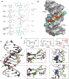
Fig. 4.. Structure of the colibactin-DNA ICL.
(A) Schematic and atom numbering of colibactin, and its hydrogen bonding and electrostatic interaction with nucleotide bases as denoted by magenta dashed lines. (B) Surface representation of the structure showing the pyrrolidine, thiazole and pyrrole rings stacked in between the minor groove edges of the DNA parallel to the long axis. (C) Portions of a 2D HSQC experiment with a [15N,13C]-colibactin-DNA ICL sample showing two hydrogen atoms correlated with N37 indicating protonation of the nitrogen (left). The equivalent N3/3’ (center) and N1/1’ (right) nitrogens and their associated protons have different chemical shifts indicating different environments and the pseudo-symmetric nature of the interaction. (D) Cartoon representation of the interaction and zoom-in views showing the placement of, i) colibactin iminium moiety with the electronegative atoms of the center T7A8 residues of the DNA (top), and ii) the amide hydrogen (H13) and the pyrrolidinone ring hydrogen (H12) interacting with T10b and A5a nucleobase (bottom). For clarity, only the inner motif residues (A6-T9) on the A-strand have been labeled.
Fig. 5.
Fig. 5.. The unstable central motif of colibactin influences its sequence specificity for ICL formation.
(A) Structural comparison of bacterially produced colibactin and a synthetic ‘stable’ colibactin analog suggests the analog may have altered specificity for ICL formation. (B) Quantification of ICL formation by co-incubation with bacteria through denaturing gel analysis for 50mers containing a double GC substitution within the sequence 5′-AATATT-3′. Results are quantified through densitometry and shown as bar plots. (C) Quantification of ICL formation by treatment with a colibactin analog through denaturing gel analysis for 50mers containing a double GC substitution within the sequence 5’-AATATT-3’. Results are quantified through densitometry and shown as bar plots. All data are mean +/− s.d. and n = 3 biological replicates. **** P < 0.0001; ** P < 0.01; * P < 0.05; not significant (ns) P > 0.05, one-way ANOVA and Tukey’s multiple comparison test.
Figure 6.
Figure 6.. Electrostatic and steric interactions involving colibactin’s unstable central motif likely drive sequence specificity for DNA alkylation.
Electrostatic potential (ESP) values of proposed colibactin structures (V in kJ·mol−1·e−1) obtained from density functional theory (DFT) optimization calculations of proposed colibactin structures at the B3LYP-D3/6-31G* level of theory. ESP values were calculated for (A) proposed structures of free colibactin (B) proposed colibactin structures crosslinked to the doubly charged DNA sequence GAATATTC (C) proposed colibactin structures crosslinked to the doubly charged double mutant DNA sequence 5′-GAACGTTC-3′. Proposed colibactin structures included the α-ketoiminium (colibactin=NH2+), α-ketoimine (colibactin=NH), diketone (colibactin=O), CH2–CH2 (colibactin–methylene), and enolamine (colibactin–NH2) central functional groups. The indices X and Y correspond to the heavy element indices of the central functional group. X=N and Y=O for colibactin=NH2+, colibactin=NH, and colibactin–NH2. X=O and Y=O for colibactin=O. X=C and Y=C for colibactin–methylene. (D–F) Electrostatic potential calculations using DNAPhi predicting the highly electronegative environment in the sequence containing the preferred AATATT motif (black) and the decrease in electronegativity upon sequence substitution in the central motif (orange). (G-I) Comparison of the NMR structure with structural modeling of the substituted sequences shown in E and F, showing potential steric clashes between colibactin and DNA (orange spheres).

Similar articles

References

    1. El Tekle G., Andreeva N., Garrett W. S., The role of the microbiome in the etiopathogenesis of colon cancer. Annu. Rev. Physiol. 86, 453–478 (2024). - PubMed
    1. White M. T., Sears C. L., The microbial landscape of colorectal cancer. Nat. Rev. Microbiol. 22, 240–254 (2024). - PubMed
    1. Dougherty M. W., Jobin C., Shining a light on colibactin biology. Toxins (Basel) 13, 346 (2021). - PMC - PubMed
    1. Wernke K. M., Xue M., Tirla A., Kim C. S., Crawford J. M., Herzon S. B., Structure and bioactivity of colibactin. Bioorg. Med. Chem. Lett. 30, 127280 (2020). - PMC - PubMed
    1. Nougayrède J.-P., Homburg S., Taieb F., Boury M., Brzuszkiewicz E., Gottschalk G., Buchrieser C., Hacker J., Dobrindt U., Oswald E., Escherichia coli induces DNA double-strand breaks in eukaryotic cells. Science 313, 848–851 (2006). - PubMed

Publication types

LinkOut - more resources