[Preprint]. 2025 May 29:2025.05.28.656696.
doi: 10.1101/2025.05.28.656696.

SLX4IP acts in parallel to FANCM to limit BLM-dependent replication stress at ALT telomeres

Affiliations

SLX4IP acts in parallel to FANCM to limit BLM-dependent replication stress at ALT telomeres

Jessica Spindler et al. bioRxiv. .

Abstract

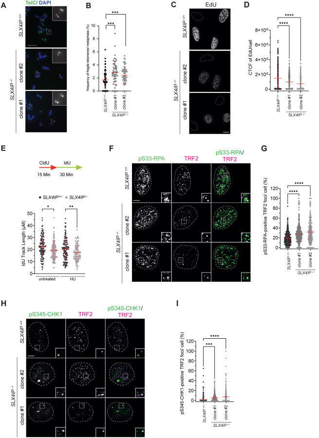
Alternative Lengthening of Telomeres (ALT) is a telomerase-independent telomere maintenance mechanism that enables cancer cells to gain unlimited replicative capacity. ALT relies on recombination-mediated telomere elongation and is promoted by telomeric replication stress. However, ALT requires strict regulation, as excessive replication stress or recombination are cytotoxic. Central to ALT is the RecQ helicase BLM, which regulates telomeric replication stress and promotes telomere recombination and DNA synthesis. Despite its key role in the ALT pathway, BLM must be tightly regulated to prevent deleterious outcomes. Here, we identify SLX4IP as a key suppressor of BLM-driven replication stress at ALT telomeres. Loss of SLX4IP in ALT-positive cells leads to BLM-dependent telomeric replication stress and impaired replication fork progression. Mechanistically, SLX4IP limits the unwinding of unligated Okazaki fragments by BLM on the lagging strand during telomere replication. This reduces the formation of toxic 5' DNA flaps and prevents hyperactivation of ATR signalling and deleterious recombination levels. We also uncover a synthetic lethal interaction between SLX4IP and FANCM, an ATPase/translocase that is a known regulator of BLM at telomeric replication forks in ALT cells. We demonstrate that SLX4IP and FANCM act in parallel to restrain BLM activity, thereby maintaining the balance of replication stress and recombination that is necessary for productive ALT. These findings reveal a vulnerability in ALT-positive cancers lacking SLX4IP and establish SLX4IP as a potential biomarker for therapeutic strategies targeting FANCM.

Keywords: ALT; BLM; FANCM; SLX4IP; cancer; genome stability; lagging-strand replication; replication stress; telomere.


Conflict of interest statement

DECLARATION OF INTERESTS The authors declare no completing interests.

Figures

Figure 1.
Figure 1.. SLX4IP depletion activates the replication stress response at ALT telomeres.
(A) U2OS cells were fixed, and metaphases were processed for telomere PNA (TelC) FISH and DAPI. Insets are 3X magnifications of the indicated fields. Scale bar represents 100 μm. Arrows indicate fragile telomeres. (B) Quantification of (A). The number of fragile telomeres counted in each metaphase was normalized to metaphases size. Data are represented as mean ± SD; n=2 with at least 30 metaphases per experiment; ***p < 0.001, one-way ANOVA. (C) U2OS cells were subjected to a 30 min EdU pulse before fixation, followed by staining of EdU via a Click-IT reaction. Dotted line represents DAPI (not shown). Scale bar represents 10 μm. (D) Quantification of (B). The EdU signal was quantified as corrected total nuclear intensity per cell. At least 100 cells per condition and experiment were counted. Data are represented as mean ± SD; n=3; ****p < 0.0001, one-way ANOVA. (E) U2OS cells were labelled with CldU and IdU and treated with 50 μM Hydroxyurea for the indicated times before DNA combing. Individual IdU fiber lengths are plotted. Median is indicated. P values were derived from ANOVA with Dunn’s multiple comparisons post-test. (F) U2OS cells were pre-extracted, fixed and processed for pSer33-RPA and TRF2 immunofluorescence. Insets are 3X magnifications of the indicated fields. Dotted line represents DAPI (not shown). Scale bar represents 5 μm. (G) Quantification of (E). The percentages of TelC foci that overlap with pSer33-RPA foci in each cell in were quantified. At least 100 cells per condition and experiment were counted. Data are represented as mean ± SD; n=3; ****p < 0.0001, one-way ANOVA. (H) U2OS cells were pre-extracted, fixed and processed for pSer345-CHK1 and TRF2 immunofluorescence. Insets are 3X magnifications of the indicated fields. Dotted line represents DAPI (not shown). Scale bar represents 5 μm. (I) Quantification of (G). The percentages of TelC foci that overlap with pSer345-CHK1 foci in each cell were quantified. At least 100 cells per condition and experiment were counted. Data are represented as mean ± SD; n=2; ****p < 0.0001, one-way ANOVA.
Figure 2
Figure 2. SLX4IP acts in parallel to FANCM to limit replication stress at ALT telomeres.
(A) U2OS Cells were transfected either with non-targeting siRNA (siCTRL) or FANCM targeting siRNA (siFANCM), pre-extracted, fixed and processed for pSer33-RPA immunofluorescence followed by telomeric PNA (TelC) FISH. Insets are 3X magnifications of the indicated fields. Dotted line represents DAPI (not shown). Scale bar represents 5 μm. (B) Quantification of (A). The percentages of TelC foci that overlap with pSer33-RPA foci in each cell were quantified. At least 100 cells per condition and experiment were counted. Data are represented as mean ± SD; n=3; ****p < 0.0001, one-way ANOVA. Knockdown validations are shown in Supplementary Figure 2A. (C) U2OS cells were transfected either with non-targeting siRNA (siCTRL) or FANCM targeting siRNA (siFANCM), pre-extracted, fixed and processed for pSer345-CHK1 immunofluorescence followed by telomeric PNA (TelC) FISH. Insets are 3X magnifications of the indicated fields. Dotted line represents DAPI (not shown). Scale bar represents 5 μm. (D) Quantification of (C). The percentages of TelC foci that overlap with pSer345-CHK1 foci in each cell were quantified. At least 80 cells per condition and experiment were counted. Data are represented as mean ± SD; n=3; ****p < 0.0001, one-way ANOVA. Knockdown validations are shown in Supplementary Figure 2B. (E) U2OS cells were transfected either with non-targeting siRNA (siCTRL) or FANCM targeting siRNA (siFANCM), pre-extracted, fixed and processed for PML immunofluorescence followed by telomeric PNA (TelC) FISH. Insets are 3X magnifications of the indicated fields. Dotted line represents DAPI (not shown). Scale bar represents 5 μm. (F) Quantification of (E). APBs were quantified as the percentage of TelC foci that overlap with PML foci in each cell. At least 95 cells per condition and experiment were counted. Data are represented as mean ± SD; n=5; ****p < 0.0001, one-way ANOVA. Knockdown validations are shown in Supplementary Figure 2C. (G) U2OS cells were transfected either with non-targeting siRNA (siCTRL) or FANCM targeting siRNA (siFANCM) and seeded in a clonogenic survival assay. (H) Quantification of (G). Data are represented as mean ± SD; n=4; ****p < 0.0001, ***p < 0.001, **p < 0.01, one-way ANOVA. Knockdown validations are shown in Supplementary Figure 2D.
Figure 3.
Figure 3.. The synthetic lethal interaction between SLX4IP and FANCM is dependent on BLM.
(A) Whole cell lysates of U2OS cells were separated by SDS-PAGE and analyzed for BLM levels by immunoblotting. Tubulin was used as loading control. The numbers on the left denote the molecular weight in kDa. (B) U2OS cells were transfected either with non-targeting siRNA (siCTRL) or FANCM targeting siRNA (siFANCM), pre-extracted, fixed and processed for BLM immunofluorescence followed by telomeric PNA (TelC) FISH. Insets are 3X magnifications of the indicated fields. Dotted line represents DAPI (not shown). Scale bar represents 5 μm. (C) Quantification of (B). The percentages of TelC foci that overlap with BLM foci in each cell were quantified. At least 95 cells per condition and experiment were counted. Data are represented as mean ± SD; n=3; ****p < 0.0001, one-way ANOVA. Knockdown validations are shown in Supplementary Figure 3A. (D) U2OS cells were transfected either with non-targeting siRNA (siCTRL) or knocked down for FANCM (siFANCM), BLM (siBLM) or both (siFANCM/siBLM). Cells were pre-extracted, fixed and processed for pSer33-RPA immunofluorescence followed by telomeric PNA (TelC) FISH. Insets are 3X magnifications of the indicated fields. Dotted line represents DAPI (not shown). Scale bar represents 5 μm. (E) Quantification of (D). The percentages of TelC foci that overlap with pSer33-RPA foci in each cell were quantified. At least 68 cells per condition and experiment were counted. Data are represented as mean ± SD; n=3; ****p < 0.0001, one-way ANOVA. Knockdown validations are shown in Supplementary Figure 3B. (F) U2OS cells were transfected either with non-targeting siRNA (siCTRL) or knocked down for FANCM (siFANCM), BLM (siBLM) or both (siFANCM/siBLM). Cells were then seeded in a clonogenic survival assay. (G) Quantification of (F). Data are represented as mean ± SD; n=4; *p < 0.05, one-way ANOVA; ns, not significant. Knockdown validations are shown in Supplementary Figure 3C.
Figure 4.
Figure 4.. SLX4IP depletion causes BLM-dependent lagging-strand replication stress.
(A) Loss of SLX4IP accumulates FEN1 and histones at replication forks. U2OS were subjected to a 20 min EdU pulse to label newly synthesized DNA, followed by iPOND coupled to mass spectrometry. The graph shows the log2 ratio intensity of proteins accumulated at replication forks in SLX4IP−/− compared to SLX4IP+/+ cells. (B) U2OS cells were labelled with CldU and IdU before DNA combing and in gel S1 nuclease treatment. Individual IdU fibre lengths are plotted. Median is indicated. P values were derived from ANOVA with Dunn’s multiple comparisons post-test. (C) U2OS cells were treated with 10 μM PARG inhibitor for 30 min, immediately fixed and processed for ADP ribose immunofluorescence. Dotted line represents DAPI (not shown). Scale bar represents 10 μm. (D) Quantification of (C). ADP ribose foci in each cell were quantified and normalized to the nucleus size. At least 100 cells per condition and experiment were counted. Data are represented as mean ± SD; n=3; ****p < 0.0001, one-way ANOVA. (E) U2OS cells were transfected either with non-targeting siRNA (siCTRL) or BLM targeting siRNA (siBLM). Cells were treated with 10 μM PARG inhibitor for 30 min, immediately fixed and processed for ADP ribose immunofluorescence. Dotted line represents DAPI (not shown). Scale bar represents 10 μm. (F) Quantification of (E). ADP ribose foci in each cell were quantified and normalized to the nucleus size. At least 100 cells per condition and experiment were counted. Data are represented as mean ± SD; n=3; ****p < 0.0001, one-way ANOVA; ns, not significant. Knockdown validations are shown in Supplementary Figure 4H.

Similar articles

References

    1. Maciejowski J. & De Lange T. Telomeres in cancer: tumour suppression and genome instability. Nat Rev Mol Cell Biol 18, 175–186 (2017). - PMC - PubMed
    1. Bryan T. M., Englezou A., Dalla-Pozza L., Dunham M. A. & Reddel R. R. Evidence for an alternative mechanism for maintaining telomere length in human tumors and tumor-derived cell lines. Nat Med 3, 1271–1274 (1997). - PubMed
    1. Henson J. D. & Reddel R. R. Assaying and investigating Alternative Lengthening of Telomeres activity in human cells and cancers. FEBS Letters 584, 3800–3811 (2010). - PubMed
    1. Dilley R. L. & Greenberg R. A. ALTernative Telomere Maintenance and Cancer. Trends in Cancer 1, 145–156 (2015). - PMC - PubMed
    1. Dilley R. L. et al. Break-induced telomere synthesis underlies alternative telomere maintenance. Nature 539, 54–58 (2016). - PMC - PubMed

Publication types

LinkOut - more resources