[Preprint]. 2025 May 27:2025.05.23.655815.
doi: 10.1101/2025.05.23.655815.

COVID-19 induces persistent transcriptional changes in adipose tissue that are not associated with Long COVID

Affiliations

COVID-19 induces persistent transcriptional changes in adipose tissue that are not associated with Long COVID

Soneida DeLine-Caballero et al. bioRxiv. .

Abstract

Long COVID is a heterogeneous condition characterized by a wide range of symptoms that persist for 90 days or more following SARS-CoV-2 infection. Now more than five years out from the onset of the SARS-CoV-2 pandemic, the mechanisms driving Long COVID are just beginning to be elucidated. Adipose tissue has been proposed as a potential reservoir for viral persistence and tissue dysfunction contributing to symptomology seen in Long COVID. To test this hypothesis, we analyzed subcutaneous adipose tissue (SAT) from two cohorts: participants with subacute COVID-19 (28-89 days post-infection) compared to pre-pandemic controls, and participants with Long COVID compared to those with those classified as "indeterminate" based on the RECOVER-Adult Long COVID Research Index (12-47 months post-infection). We found no evidence of persistent SARS-CoV-2 RNA in adipose tissue in any participant. SAT from participants with subacute COVID-19 displayed significant transcriptional remodeling, including depleted immune activation pathways and upregulated Hox genes and integrin interactions, suggesting resident immune cell exhaustion and perturbations in tissue function. However, no consistent changes in gene expression were observed between Long COVID samples and samples from indeterminant participants. Thus, SAT may contribute to inflammatory dysregulation following COVID-19, but does not appear to play a clear role in Long COVID pathophysiology. Further research is needed to clarify the role of adipose tissue in COVID-19 recovery.

Keywords: Long COVID; RNA Sequencing; SARS-CoV-2; Subacute COVID-19; Subcutaneous Adipose Tissue; Transcriptome.


Conflict of interest statement

Conflict of Interest JZL consults for Merck. CAB consults for Immunebridge on topics unrelated to the current research. All other authors declare that the research was conducted in the absence of any commercial or financial relationships that could be construed as a potential conflict of interest.

Figures
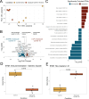
Figure 1
Figure 1. Transcriptional changes in SAT from participants with subacute COVID-19.
(A) Dimensionality reduction by principal components (PC) analysis on all genes for participant samples in cohort 1 (subacute COVID-19 and pre-pandemic participant samples). Each data point corresponds to the SAT sample of a participant. PC1, principal component 1; PC2, principal component 2; percentage expresses contribution to the overall data variability (B) Volcano plot visualization of the differentially expressed genes between subacute COVID-19 participant and pre-pandemic control samples. Horizontal dashed line represents padj = 0.05 and vertical dashed lines represent an absolute log fold change of 1. (C) Significantly enriched (padj < 0.0001) Blood Transcriptional Modules (BTM) within the upregulated and downregulated differentially expressed genes. (D) Box plot of Immune activation gene scores for subacute COVID-19 samples (n=5) and pre-pandemic control samples, (n=4); p-values generated with Wilcoxon rank sum test. (E) Box plot Hox Cluster III BTM Gene Scores for subacute COVID-19 participants (n=5) and pre-pandemic control participants (n=5); p-values generated by Wilcoxon rank sum test. For boxplot visualizations, the top box line indicates the 75th quartile gene score; middle box line indicates the mean gene score, and the bottom box line represents the 25th quartile gene score
Figure 2
Figure 2. Expression of Immune Activation and Hox Cluster BTMs Are Strongly Associated with Resident Tissue Macrophages and Preadipocytes in SAT.
(A) Uniform Manifold Approximation and Projection (UMAP) of Single Cell RNA (scRNA) dataset from Stromal Vascular Fraction of human SAT (GSE208034) colored by cell type (annotated using Seurat) (B-C) UMAP from A, colored by gene score generated on the expression of genes within the Blood Transcription Modules (BTMs) of (B) immune activation and (C) Hox clusters I-VI.
Figure 3
Figure 3. Long COVID is not associated with significant transcriptional changes compared to those with Indeterminate symptoms.
(A) Dimensionality reduction by principal components analysis based on log2 normalized counts of all genes for participant samples in cohort 2 (Long COVID and indeterminate Status participant samples). Each data point corresponds to the sample of one participant. PC1, principal component 1; PC2, principal component 2; percentage expresses contribution to the overall data variability (B) Volcano plot visualization of the differentially expressed genes between Long COVID participants and indeterminate control samples. Horizontal dashed line represents padj = 0.05 and vertical dashed lines represent an absolute log fold change of 1. (C-D) Wilcoxon rank-sum p-values comparing gene scores between Control versus Long COVID-19 samples. For boxplot visualizations, the top box line indicates the 75th quartile gene score; middle box line indicates the mean gene score, and the bottom box line represents the 25th quartile gene score.

Similar articles

  • Antibody tests for identification of current and past infection with SARS-CoV-2.
    Fox T, Geppert J, Dinnes J, Scandrett K, Bigio J, Sulis G, Hettiarachchi D, Mathangasinghe Y, Weeratunga P, Wickramasinghe D, Bergman H, Buckley BS, Probyn K, Sguassero Y, Davenport C, Cunningham J, Dittrich S, Emperador D, Hooft L, Leeflang MM, McInnes MD, Spijker R, Struyf T, Van den Bruel A, Verbakel JY, Takwoingi Y, Taylor-Phillips S, Deeks JJ; Cochrane COVID-19 Diagnostic Test Accuracy Group. Fox T, et al. Cochrane Database Syst Rev. 2022 Nov 17;11(11):CD013652. doi: 10.1002/14651858.CD013652.pub2. Cochrane Database Syst Rev. 2022. PMID: 36394900 Free PMC article.
  • Rapid, point-of-care antigen tests for diagnosis of SARS-CoV-2 infection.
    Dinnes J, Sharma P, Berhane S, van Wyk SS, Nyaaba N, Domen J, Taylor M, Cunningham J, Davenport C, Dittrich S, Emperador D, Hooft L, Leeflang MM, McInnes MD, Spijker R, Verbakel JY, Takwoingi Y, Taylor-Phillips S, Van den Bruel A, Deeks JJ; Cochrane COVID-19 Diagnostic Test Accuracy Group. Dinnes J, et al. Cochrane Database Syst Rev. 2022 Jul 22;7(7):CD013705. doi: 10.1002/14651858.CD013705.pub3. Cochrane Database Syst Rev. 2022. PMID: 35866452 Free PMC article.
  • Signs and symptoms to determine if a patient presenting in primary care or hospital outpatient settings has COVID-19.
    Struyf T, Deeks JJ, Dinnes J, Takwoingi Y, Davenport C, Leeflang MM, Spijker R, Hooft L, Emperador D, Domen J, Tans A, Janssens S, Wickramasinghe D, Lannoy V, Horn SRA, Van den Bruel A; Cochrane COVID-19 Diagnostic Test Accuracy Group. Struyf T, et al. Cochrane Database Syst Rev. 2022 May 20;5(5):CD013665. doi: 10.1002/14651858.CD013665.pub3. Cochrane Database Syst Rev. 2022. PMID: 35593186 Free PMC article.
  • SARS-CoV-2-neutralising monoclonal antibodies for treatment of COVID-19.
    Kreuzberger N, Hirsch C, Chai KL, Tomlinson E, Khosravi Z, Popp M, Neidhardt M, Piechotta V, Salomon S, Valk SJ, Monsef I, Schmaderer C, Wood EM, So-Osman C, Roberts DJ, McQuilten Z, Estcourt LJ, Skoetz N. Kreuzberger N, et al. Cochrane Database Syst Rev. 2021 Sep 2;9(9):CD013825. doi: 10.1002/14651858.CD013825.pub2. Cochrane Database Syst Rev. 2021. PMID: 34473343 Free PMC article.
  • Workplace interventions to reduce the risk of SARS-CoV-2 infection outside of healthcare settings.
    Pizarro AB, Persad E, Durao S, Nussbaumer-Streit B, Engela-Volker JS, McElvenny D, Rhodes S, Stocking K, Fletcher T, Martin C, Noertjojo K, Sampson O, Verbeek JH, Jørgensen KJ, Bruschettini M. Pizarro AB, et al. Cochrane Database Syst Rev. 2022 May 6;5(5):CD015112. doi: 10.1002/14651858.CD015112.pub2. Cochrane Database Syst Rev. 2022. Update in: Cochrane Database Syst Rev. 2024 Apr 10;4:CD015112. doi: 10.1002/14651858.CD015112.pub3. PMID: 35514111 Free PMC article. Updated.

References

    1. Skevaki C, Moschopoulos CD, Fragkou PC, Grote K, Schieffer E, Schieffer B. Long COVID: Pathophysiology, current concepts, and future directions. J Allergy Clin Immunol (2025) 155:1059–1070. doi: 10.1016/j.jaci.2024.12.1074 - DOI - PubMed
    1. WHO Coronavirus Disease (COVID-19) Dashboard. Bangladesh Physiother J (2020) 10: doi: 10.46945/bpj.10.1.03.01 - DOI
    1. Adjaye-Gbewonyo D, Vahratian A, Perrine CG, Bertolli J. Long COVID in Adults: United States, 2022. NCHS data brief (2023)1–8. - PubMed
    1. Hamlin RE, Blish CA. Challenges and opportunities in long COVID research. Immunity (2024) 57:1195–1214. doi: 10.1016/j.immuni.2024.05.010 - DOI - PMC - PubMed
    1. Al-Aly Z, Davis H, McCorkell L, Soares L, Wulf-Hanson S, Iwasaki A, Topol EJ. Long COVID science, research and policy. Nat Med (2024) 30:2148–2164. doi: 10.1038/s41591-024-03173-6 - DOI - PubMed

Publication types