[Preprint]. 2025 Jun 3:2025.06.02.655683.
doi: 10.1101/2025.06.02.655683.

Structural basis of liver de-targeting and neuronal tropism of CNS-targeted AAV capsids

Affiliations

Structural basis of liver de-targeting and neuronal tropism of CNS-targeted AAV capsids

Tyler J Brittain et al. bioRxiv. .

Abstract

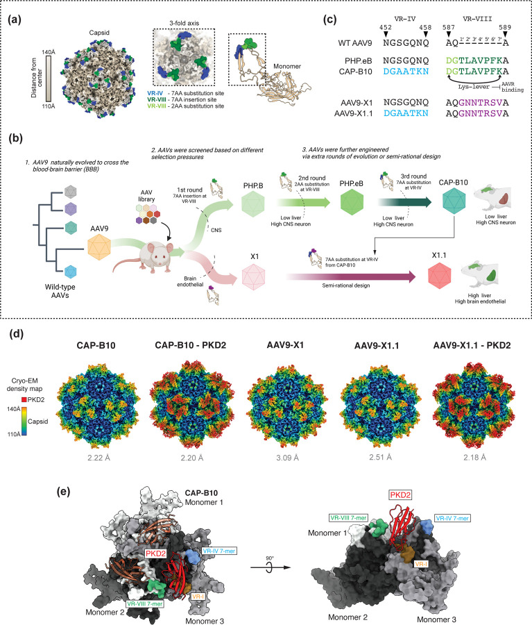
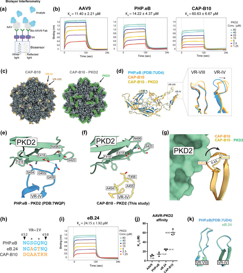
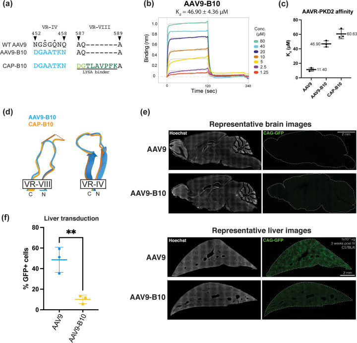
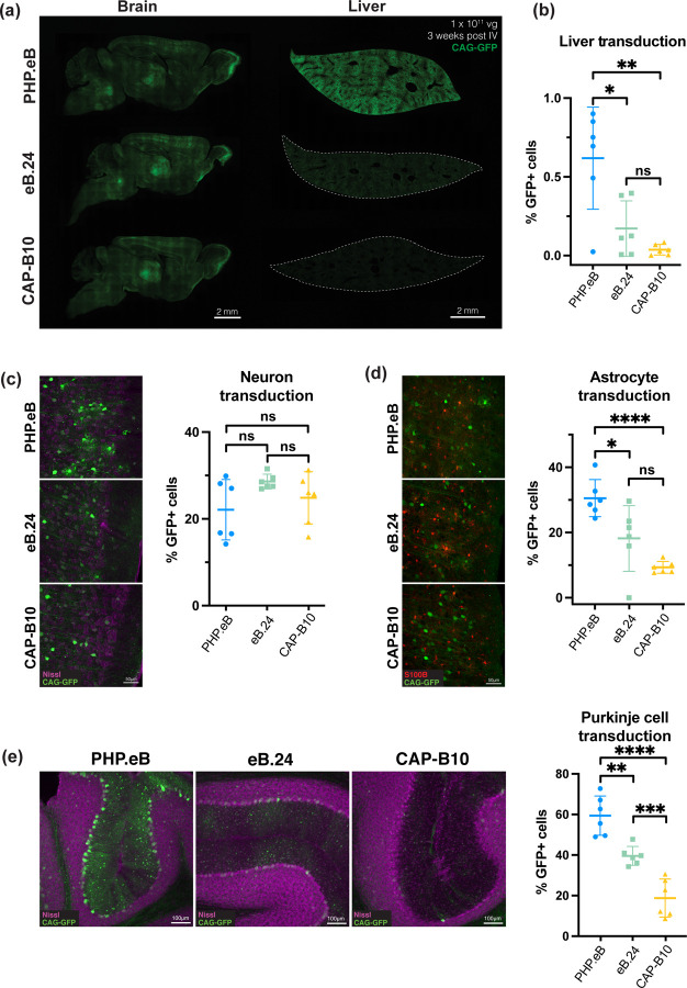
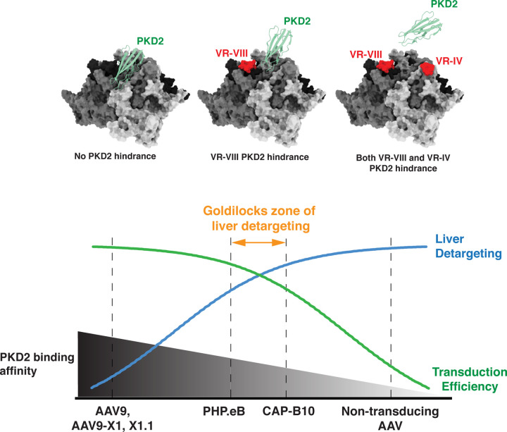
Crossing the blood-brain barrier while minimizing liver transduction is a key challenge in developing safe adeno-associated virus (AAV) vectors for treating brain disorders. In mice, the engineered capsid PHP.eB shows enhanced brain transduction, while the further engineered CAP-B10 is also de-targeted from astrocytes and liver. Here, we solve cryo-EM structures of CAP-B10 and its complex with AAV receptor (AAVR) domain PKD2, at 2.22 and 2.20 Å resolutions, respectively. These structures reveal a structural motif that hinders AAVR binding, which we confirm by measuring affinities. We show that this motif is transferable to other capsids by solving cryo-EM structures of AAV9-X1 and AAV9-X1.1, without and with PKD2, at 3.09, 2.51, and 2.18 Å, respectively. Using this structural information, we designed and validated novel AAV variants with reduced liver and altered brain cell tropism in vivo. Overall, our findings demonstrate that rationally modulating AAVR affinity can alter liver targeting and cellular tropism.


Conflict of interest statement

Competing interests V.G. is a cofounder and board member of Capsida Biotherapeutics, a fully integrated AAV engineering and gene therapy company. The remaining authors declare no competing interests.

Figures

Figure 1:
Figure 1:. Structure of engineered CNS-enhanced AAVs.
(a) Location of engineered sites on the AAV capsid shown on structure of PHP.eB (PDB: 7UD4). (b) Schematic of directed evolution efforts yielding capsids with high transduction of rodent CNS and low transduction of liver (PHP.B, PHP.eB, and CAP-B10; green arrows), and high transduction of brain endothelial cells and liver (AAV9-X1 and AAV9-X1.1; red arrows). (c) Sequence alignment of VR-IV and VR-VIII regions in engineered capsids. The CAP-B10 7-mer substitution is shown in blue, the PHP.eB 7-mer insertion in green, two point mutations in light green, and the AAV9-X1 7-mer insertion in purple. D587 and K7’ in PHP.eB and CAP-B10, form the “lysine-lever” which diminishes AAVR-PKD2 binding. (d) Single-particle cryo-EM maps of engineered capsids with and without PKD2, the AAV9 binding domain of AAVR. In the maps, color indicates distance from the center of the capsid. (e) Binding interface between PKD2 and 3-fold face of AAV. AAVR-PKD2 is red, VR-I is orange, 7-mer substitution in VR-IV is blue, 7-mer insertion in VR-VIII is green. The individual monomers of the AAV 3-fold face are colored white, light gray, and dark gray.
Figure 2:
Figure 2:. VR-IV modification in CAP-B10 reduces AAVR-PKD2 affinity.
(a) Schematic of biolayer interferometry (BLI) to measure the binding affinity between AAV capsids and AAVR-PKD2 domain. Biotinylated AAV9 affinity ligand is immobilized on a streptavidin-coated biosensor, AAVs are loaded as the analyte, and binding is quantified by sensorgrams. (b) Representative sensorgrams detail the interactions between AAV9, PHP.eB, and CAP-B10 with AAVR-PKD2. Assays were performed three times, with one representative dataset shown. The averaged equilibrium dissociation constant (Kd) is presented for each capsid. (c) Cryo-EM maps of CAP-B10 without and with PKD2. In the uncomplexed map, VR-IV density is shown in orange and VR-VIII density in brown. In the complex map, PKD2 density is shown in green. (d) Atomic models of the VP3 subunit of PHP.eB (PDB: 7UD4), CAP-B10 (this study), and CAP-B10 complexed with PKD2 (this study), overlaid to show structural differences and similarities. Inset shows (left) that the structure of VR-VIII in PHP.eB and CAP-B10 is similar, and in the presence of PKD2 additional residues become resolvable, and (right) VR-IV loop structural differences between CAP-B10 (orange) and PHP.eB (blue). Arrow indicates the lower position of VR-IV in CAP-B10 relative to PHP.eB, and the additional conformational change when CAP-B10 is bound to PKD2 (yellow). (e) PHP.eB has favorable amino acids at positions 456 and 454 to interact with PKD2. Q456 on PHP.eB can form a hydrogen bond to PKD2 E418 and N496, with PKD2 S420 and PHP.eB S454 aiding in creating a hydrophilic environment. Bulky F416 on PKD2 is positioned away from these hydrophilic residues thereby preventing steric clashes. Dashed sky blue line indicates potential hydrogen bonding (f) Our CAP-B10 PKD2 complex structure reveals that two key mutations, S454A and Q456T, play a key role in disrupting the PKD2 interaction. Short T456 prevents hydrophilic interaction with E418, N496, and S420 on PKD2. A454 sterically clashes with F416 on PKD2, causing the VR-IV loop to bend away from PKD2. In addition to these two residues, the generally downwardly bent structure of VR-IV further distances T456 from hydrophilic PKD2 residues. (g) Comparison of structures of CAP-B10 (orange) and CAP-B10 complexed with PKD2 (yellow) shows that PKD2 induces a conformational change in the VR-IV of CAP-B10. The greatest shift occurs at A454, which is shifted 6.4Å farther away from PKD2. (h) VR-IV sequences of PHP.eB, eB.24, and CAP-B10. The two point mutations in eB.24 are highlighted in orange, S454A and Q456T. (i) Representative BLI sensorgram of eB.24 binding of AAVR-PKD2. (j) Affinity values of engineered capsids for AAVR-PKD2. Kd values are an average of 3 replicates. (k) Cryo-EM-based atomic model of eB.24 overlaid with the structure of PHP.eB (PDB: 7UD4). The two point mutations (S454A, Q456T) in eB.24 do not modify the VR-IV backbone structure of PHP.eB.
Figure 3:
Figure 3:. Structural and biophysical characterization of AAV9-X1 and AAV9-X1.1 binding to AAVR-PKD2
(a) Representative BLI sensorgrams showing the interaction of AAV9-X1 and AAV9-X1.1 with AAVR-PKD2. Kd values are averages of three replicates. (b) Comparison of AAVR-PKD2 binding affinity of AAV9, AAV9-X1, and AAV9-X1.1. AAV9-X1.1 shows weaker AAVR-PKD2 binding affinity compared to AAV9-X1, indicating that the CAP-B10 VR-IV structural motif introduced in AAV9-X1.1 reduces AAVR-PKD2 binding, similarly to CAP-B10. Note that the AAV9 - AAVR-PKD2 affinity shown here is from the same experiment as in Figure 2j. (c) Cryo-EM maps of AAV9-X1.1 in its unbound and AAVR-PKD2 bound states. In the uncomplexed structure, the VR-IV is shown in orange and VR-VIII is in brown. In the complex map, PKD2 density is highlighted in green. (d) Atomic model of VR-IV in AAV9-X1.1 (blue) complexed with PKD2 (green). As in CAP-B10, T456 loses its hydrogen bond to PKD2 E418, while A454 is pushed away due to a steric clash with PKD2 F416. (e) Atomic model of VR-VIII in AAV9-X1.1 (blue) complexed with PKD2 (green surface), overlaid with AAV9-X1.1 (yellow) and CAP-B10 (orange). The schematics below illustrate the interactions between PKD2 and VR-VIII of AAV9-X1.1. Left: Interaction of VR-VIII with the anterior PKD2. Right: Interaction of VR-VIII with the posterior PKD2. Asterisks denote the four polar-basic amino acids (2’NNTR5’) at the protruding end of the X1 7-mer that potentially interact with the polar-acidic surface of PKD2.
Figure 4:
Figure 4:. B10 structural motif can reduce liver transduction without brain sequestration.
(a) Sequence alignment of VR-IV and VR-VIII in AAV9, AAV9-B10, and CAP-B10. AAV9-B10 contains the B10 7-mer substitution in VR-IV but lacks the PHP.eB 7-mer in VR-VIII, preventing interaction with LY6A. (b) Representative BLI sensorgram of AAV9-B10 binding to AAVR-PKD2. The averaged Kd from triplicate measurements is shown above. (c) Comparison of the AAVR-PKD2 affinity values for AAV9, AAV9-B10, and CAP-B10. AAV9 and CAP-B10 data are repeated from Figure 2b and 2j for reference. (d) Cryo-EM structure of AAV9-B10, demonstrating that its VR-IV conformation is identical to that of CAP-B10. Additionally, AAV9’s native VR-VIII structure is not altered by the addition of the B10 structural motif at VR-IV. (e) Representative images of the brain and liver of mice following systemic delivery of AAV9 or AAV9-B10 packaging eGFP under the control of the CAG promoter. AAVs were retro-orbitally administered to the mice at a dose of 1×10¹¹ vg per animal (n = 6 animals per capsid). eGFP expression was analyzed 3 weeks post-injection. Scale bars represent 2 mm. (f) Percentage of eGFP-expressing cells in the liver of mice receiving AAV9 or AAV9-B10. AAV9-B10 has markedly decreased liver transduction compared to AAV9 (**p < 0.01). Statistical significance was determined using a two-tailed unpaired t-test.
Figure 5:
Figure 5:. AAVR-PKD2 affinity modulates liver and brain tropism.
(a) Representative fluorescence images showing eGFP expression in the brain and liver following retro-orbital delivery of PHP.eB, eB.24, or CAP-B10. AAVs packaging the fluorescent reporter eGFP under the ubiquitous CAG promoter were administered retro-orbitally at a dose of 1×10¹¹ vg per mouse (n = 6 animals per capsid). eGFP expression was analyzed 3 weeks post-injection. The fluorescent reporter was used to quantify varying levels of brain and liver transduction. Scale bars represent 2 mm. (b) Quantification of eGFP-positive cells in the liver shows a correlation with the binding affinity for AAVR-PKD2. These findings demonstrate that liver tropism can be modulated by tuning AAVR-PKD2 affinity. (c) Representative images of eGFP-expressing cells co-stained with Nissl, a marker for neurons, to assess neuronal transduction in the cortex. The percentage of GFP+ neurons is consistent between PHP.eB, eB.24, and CAP-B10, suggesting that AAVR affinity does not significantly alter neuronal transduction levels. Scale bar represents 50 µm. (d) Percentage of eGFP-expressing cells co-localizing with S100B, indicating astrocyte transduction efficiency. Similar to the liver, astrocytic tropism decreases as AAVR-PKD2 affinity decreases, highlighting the role of AAVR binding in regulating astrocyte transduction. (e) Left: representative images of eGFP expression in the cerebellum delivered by PHP.eB, eB.24, or CAP-B10 where eGFP is in green and Nissl-stained neurons are in magenta. Right: percentage of eGFP-positive Purkinje cells in the cerebellum. As AAVR-PKD2 affinity decreases the transduction level of Purkinje cells decreases. Statistical significance was determined using a two-tailed unpaired t-test. *p < 0.05, **p < 0.01, and ***p < 0.001.
Figure 6:
Figure 6:. Engineering liver de-targeted AAVs requires a balanced approach.
While higher AAVR-PKD2 affinity generally enhances transduction across various organs, it also increases liver transduction. Conversely, AAVs that completely lose AAVR-PKD2 binding fail to transduce any cells. Additionally, even with reduced AAVR-PKD2 affinity, an AAV may still transduce the liver if it engages an alternative liver receptor, such as LRP6 for AAV9-X1.1. Therefore, designing liver de-targeted AAVs requires optimizing AAVR-PKD2 affinity in the context of target-specific receptor interactions.

References

    1. Ling Q., Herstine J. A., Bradbury A. & Gray S. J. AAV-based in vivo gene therapy for neurological disorders. Nat. Rev. Drug Discov. 22, 789–806 (2023). - PubMed
    1. Haery L. et al. Adeno-Associated Virus Technologies and Methods for Targeted Neuronal Manipulation. Front. Neuroanat. 13, (2019). - PMC - PubMed
    1. Verdera H. C., Kuranda K. & Mingozzi F. AAV Vector Immunogenicity in Humans: A Long Journey to Successful Gene Transfer. Mol. Ther. 28, 723–746 (2020). - PMC - PubMed
    1. Al-Zaidy S. A. & Mendell J. R. From Clinical Trials to Clinical Practice: Practical Considerations for Gene Replacement Therapy in SMA Type 1. Pediatr. Neurol. 100, 3–11 (2019). - PubMed
    1. Kang L. et al. AAV vectors applied to the treatment of CNS disorders: Clinical status and challenges. J. Controlled Release 355, 458–473 (2023). - PubMed

Publication types

LinkOut - more resources