[Preprint]. 2025 Jun 3:2025.05.30.657094.
doi: 10.1101/2025.05.30.657094.

Klf4-dependent pivotal role of smooth muscle-derived Gli1 + lineage progenitor cells in lung cancer progression

Affiliations

Klf4-dependent pivotal role of smooth muscle-derived Gli1 + lineage progenitor cells in lung cancer progression

Sizhao Lu et al. bioRxiv. .

Abstract

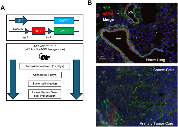
Lung cancer remains the leading cause of cancer-related deaths worldwide. Despite the remarkable efficacy of immune checkpoint inhibitors, only a subset of lung adenocarcinoma (LUAD) patients respond and many eventually progress, underscoring the need to develop novel combination therapies. The interaction between cancer cells and the tumor microenvironment (TME), a complex niche including cells of the innate and adaptive immune system, cancer-associated fibroblasts (CAFs), vascular cells, and extracellular matrix (ECM) plays a vital role in tumorigenesis and cancer progression. Using fate mapping, we previously identified a novel subpopulation of resident vascular stem cells derived from vascular smooth muscle cells, designated AdvSca1-SM (vascular Adventitial location, Stem cell antigen-1 expression, SMooth muscle origin) cells. AdvSca1-SM cells are the predominant cell type responding to vessel wall dysfunction and their selective differentiation to myofibroblasts promotes vascular fibrosis. SMC-to-AdvSca1-SM cell reprogramming is dependent on SMC induction of the transcription factor KLF4, and KLF4 is essential for maintenance of the stem cell phenotype. The function of AdvSca1-SM cells in LUAD tumorigenesis has not been explored. Using an orthotopic immunocompetent mouse model of LUAD, we demonstrate that AdvSca1-SM cells are a major component of lung tumors, significantly contributing to cancer associated fibroblasts (CAFs). Compared to AdvSca1-SM cells, CAFs have altered ECM gene expression with a reduction of a stemness signature. AdvSca1-SM-specific genetic ablation of Klf4 altered their phenotype resulting in inhibition of communication between cancer cells and CAFs, decreases in innate immunosuppressive populations, and increased T cell infiltration into the tumor. Targeting this population may represent a novel strategy to improve the response to immunotherapy.


Figures

Figure 1.
Figure 1.. AdvSca1-SM cells reside in the lung and contribute to lung cancer progression and metastasis.
(A) Schematic illustrating the experimental design. Gli1-CreERT-YFP mice were treated with tamoxifen to induce YFP gene knock-in specifically in AdvSca1-SM cells. After a washout period, mice were injected with LLC cancer cells orthotopically into the left lobe of the lung. (B) Lung tissues from naïve non-tumor-bearing (top) and LLC tumor-bearing mice (bottom) were immunofluorescently stained for AdvSca1-SM-derived cells (YFP, green), alpha smooth muscle actin (aSMA, red) for vascular and airway smooth muscle, and DAPI to identify cell nuclei. PA = pulmonary artery; Aw = airway. Representative 40x images shown from N=6.
Figure 2.
Figure 2.. AdvSca1-SM lineage cells predominately contribute to cancer-associated fibroblasts (CAFs) in the tumor microenvironment (TME).
Single cell RNA-sequencing (scRNA-Seq) was performed with single cell suspensions from naïve and LLC tumor-bearing lungs harvested from Gli1-CreERT-YFP AdvSca1-SM lineage mice. YFP+ AdvSca1-SM lineage cells and YFP cells were separated using FACS and subjected to scRNA-seq. UMAPs with annotated cell clusters (A), highlighting YFP+ samples in green (B), and separating naïve and tumor samples (C) are shown. (D) Differential gene expression (DGE) analysis was performed between YFP+ CAFs (red circle, panels B&C) and AdvSca1-SM cells (green circle, panels B&C). The results are shown in the volcano plot with select genes labeled. Positive logFC indicate up-regulation in CAFs. Pathway enrichment for up-regulated genes (E) and down-regulated genes (F) in CAFs are shown.
Figure 3.
Figure 3.. Klf4 depletion in Gli1+ AdvSca1-SM lineage cells blunts tumorigenesis and alters CAF phenotype in the TME.
Tamoxifen-treated Gli1-CreERT-YFP (WT) and Gli1-CreERT-YFP;Klf4flox/flox (Klf4 KO) mice were subjected to orthotopic LLC cell injection. Tumor volumes were measured by caliper (A) and H&E images (4x) of representative tumors from WT (left) and Klf4 KO (right) mice are shown (B) scRNA-seq was performed with tumor bearing lungs from Klf4 KO mice and the data integrated with WT scRNA-seq data. UMAP plots with annotated clusters (C), highlighting YFP+ cells in green (D), and separating WT and KO cells (E) are shown. (F) DGE analysis was performs comparing WT and Klf4 KO YFP+ CAFs. Volcano plot shown with select genes labeled. Positive logFC indicate up-regulation in Klf4 KO. Pathway enrichment results with up-regulated genes (G) and down-regulated genes (H) in Klf4 KO CAFs are shown. (I) Transcription factors (TFs) activities were inferred based on the differential gene expression analysis. Barplot showing top activated (red) TFs.
Figure 4.
Figure 4.. LLC tumor cells in Klf4 KO lung are less proliferative and more immune active.
RNA velocity analysis was performed for LLC cells in the scRNA-seq data. (A) RNA velocity is shown with stream plot. Arrows represent RNA velocity vectors, indicating the predicted future state of each cell. The length and direction of the arrows suggest the rate and trajectory of transcriptional changes, respectively. (B) LLC cells were scored for marker genes of S and G2M phase. UMAP colored by respective score are shown. (C) Composition analysis was performed to contrast LLC phenotype changes between WT and Klf4 KO lungs. (D) DGE analysis was performed between LLC cells in WT and Klf4 KO samples. Volcano plot with select genes labeled. Positive logFC indicate up-regulation in Klf4 KO. Pathway enrichment with up-regulated genes (E) and down-regulated genes (F) in Klf4 KO are shown. (G) Transcription factor (TFs) activities were inferred based on the differential gene expression analysis. Barplot shows activated (red) and suppressed (blue) TFs in LLCs from Klf4 KO lungs.
Figure 5.
Figure 5.. Increased T cell recruitment in primary tumors of AdvSca1-SM Klf4 KO mice compared to WT mice.
(A). Lung sections from LLC tumor-bearing Klf4 WT (left panels) and KO mice (right panels) were stained for CD3 (cyan), YFP (green; AdvSca1-SM lineage), aSMA (red), and DAPI (blue, nuclei) to identify recruited tumor-infiltrating T cells. Representative 40x images of CD3+ T cells in both the core and edge of tumors is shown (B) Cell count quantification revealed increased numbers of recruited T cells both in the core of the primary tumor as well as around the edges of the primary tumor. Arrows – representative CD3+ T cells. N=6; one-way ANOVA with Bonferroni’s post-hoc test; *p<0.05; **p<0.01; ***p<0.001.
Figure 6.
Figure 6.. Cell-cell communication analysis reveals Klf4 depletion in Gli1+ AdvSca1-SM lineage cells renders AdvSca1-SM-derived CAFs unresponsive to signaling from LLC cells.
(A) Steady-state ligand-receptor inference and intercellular context factorization (ICF) was performed using LIANA and Tensor-cell2cell. Cell-cell communication was factorized into 9 factors, and their content loading were plotted between WT naïve, WT Tumor, and Klf4 KO tumor conditions. Differences between groups was examined using independent t-test with false discovery rate correction (Benjamini-Hochberg method), as implemented by Tensor-cell2cell python package. ns not significant, *p<0.05, **p<0.01 Heatmaps depicting the sender clusters (B) and receiver clusters (C) loadings across intercellular communication factors is shown.

References

    1. Siegel R.L., Miller K.D., and Jemal A. (2020). Cancer statistics, 2020. CA Cancer J Clin 70, 7–30. 10.3322/caac.21590. - DOI - PubMed
    1. Reck M., Rodríguez-Abreu D., Robinson A.G., Hui R., Csőszi T., Fülöp A., Gottfried M., Peled N., Tafreshi A., Cuffe S., et al. (2016). Pembrolizumab versus Chemotherapy for PD-L1-Positive Non-Small-Cell Lung Cancer. N Engl J Med 375, 1823–1833. 10.1056/NEJMoa1606774. - DOI - PubMed
    1. Sezer A., Kilickap S., Gümüş M., Bondarenko I., Özgüroğlu M., Gogishvili M., Turk H.M., Cicin I., Bentsion D., Gladkov O., et al. (2021). Cemiplimab monotherapy for first-line treatment of advanced non-small-cell lung cancer with PD-L1 of at least 50%: a multicentre, open-label, global, phase 3, randomised, controlled trial. Lancet 397, 592–604. 10.1016/S0140-6736(21)00228-2. - DOI - PubMed
    1. Gandhi L., Rodríguez-Abreu D., Gadgeel S., Esteban E., Felip E., De Angelis F., Domine M., Clingan P., Hochmair M.J., Powell S.F., et al. (2018). Pembrolizumab plus Chemotherapy in Metastatic Non-Small-Cell Lung Cancer. N Engl J Med 378, 2078–2092. 10.1056/NEJMoa1801005. - DOI - PubMed
    1. Gogishvili M., Melkadze T., Makharadze T., Giorgadze D., Dvorkin M., Penkov K., Laktionov K., Nemsadze G., Nechaeva M., Rozhkova I., et al. (2022). Cemiplimab plus chemotherapy versus chemotherapy alone in non-small cell lung cancer: a randomized, controlled, double-blind phase 3 trial. Nat Med 28, 2374–2380. 10.1038/s41591-022-01977-y. - DOI - PMC - PubMed

Publication types

LinkOut - more resources