[Preprint]. 2025 Jun 6:2025.06.02.657515.
doi: 10.1101/2025.06.02.657515.

An improved model for prediction of de novo designed proteins with diverse geometries

Affiliations

An improved model for prediction of de novo designed proteins with diverse geometries

Benjamin Orr et al. bioRxiv. .

Abstract

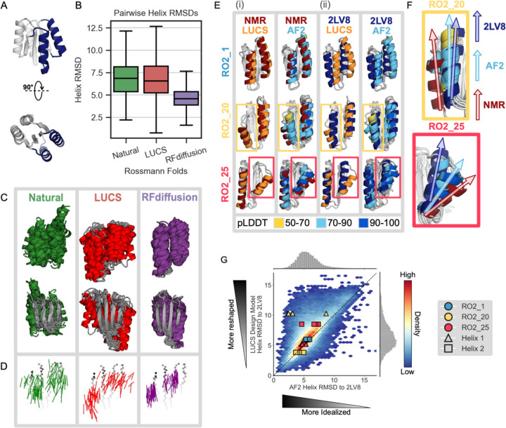
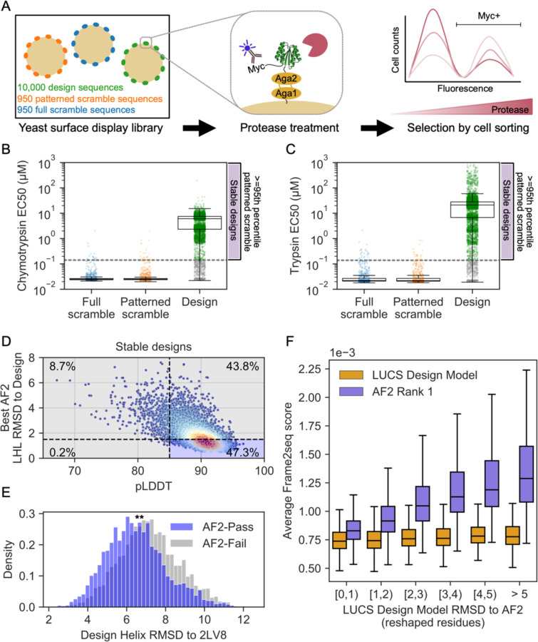
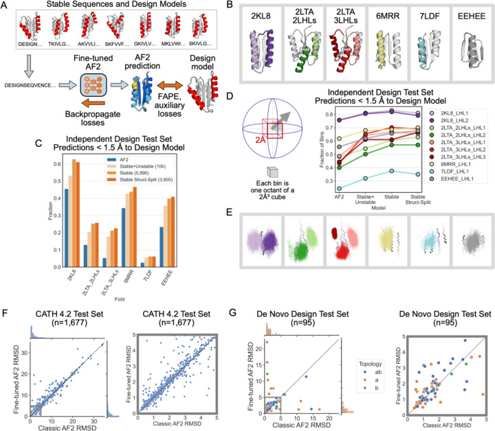
Nature uses structural variations on protein folds to fine-tune the geometries of proteins for diverse functions, yet deep learning-based de novo protein design methods generate highly regular, idealized protein fold geometries that fail to capture natural diversity. Here, using physics-based design methods, we generated and experimentally validated a dataset of 5,996 stable, de novo designed proteins with diverse non-ideal geometries. We show that deep learning-based structure prediction methods applied to this set have a systematic bias towards idealized geometries. To address this problem, we present a fine-tuned version of Alphafold2 that is capable of recapitulating geometric diversity and generalizes to a new dataset of thousands of geometrically diverse de novo proteins from 5 fold families unseen in fine-tuning. Our results suggest that current deep learning-based structure prediction methods do not capture some of the physics that underlie the specific conformational preferences of proteins designed de novo and observed in nature. Ultimately, approaches such as ours and further informative datasets should lead to improved models that reflect more of the physical principles of atomic packing and hydrogen bonding interactions and enable improved generalization to more challenging design problems.


Conflict of interest statement

Competing interests: Authors declare that they have no competing interests.

Figures

Figure 1.
Figure 1.. Deep learning-based protein structure prediction and design models predict and generate more idealized geometries than those occurring in nature or in de novo proteins designed using physics-based methods.
(A) Idealized Rossmann fold (PDB ID 2LV8) with loop-helix-loop (LHL) units diversified in this study shown in dark blue. (B) Comparison of geometric variation within groups of Rossmann fold backbones. Natural (green): 44 representative natural Rossmann fold proteins. LUCS (red): 5,996 backbones generated with loop-helix-loop unit combinatorial sampling. These designed backbones were experimentally determined to fold into stable proteins (Fig. 2). RFdiffusion (purple): 5,996 backbones generated with RFdiffusion (Methods). Boxes show the lower and upper quartiles, whiskers extend to points within 1.5 interquartile ranges of the lower and upper quartiles, and lines indicate the median values (outliers not shown). (C) Structural overlay of 44 examples from each Rossmann fold group. The 44 LUCS and RFdiffusion examples were selected by sorting backbones by their helix RMSDs to an idealized Rossmann fold (2LV8), then sampling at even intervals to span the helix RMSD range. (D) Vector representations of helices to depict geometric diversity, with the underlying beta-sheet shown in grey. Vectors indicate the helix centroids (vector tails) and average carbonyl C-O direction. (E) Structural comparisons for three experimentally validated LUCS designs (RO2_1, RO2_20 and RO2_25 from ref.) with reshaped helices shown in color: LUCS design models (orange) and 5 AF2 predictions (colored by pLDDT) are compared to (i) their experimentally-determined lowest energy NMR structures (dark red, showing close agreement with LUCS), and (ii) an idealized 2×2 Rossmann fold de novo designed protein (dark blue; PDB ID: 2LV8). (F) Closeup of comparison for non-idealized helices in RO2_20 and RO2_25, showing that AF2 predicts helix geometries closer to the idealized structure than the experimentally determined structure. (G) Comparison of AF2 structure predictions to the LUCS design models for 10,000 LUCS backbones with sequences designed with ProteinMPNN. AF2 predicts 75.8% of reshaped helices to be closer to the idealized helices in 2LV8 than to the LUCS design model (density above the diagonal). Overlaid points show the helix RMSDs to 2LV8 for the design models and AF2 predictions for the three designs with experimentally determined structures in (E) and (F).
Figure 2.
Figure 2.. AF2 fails to capture geometric diversity in a large dataset of ~6000 experimentally tested, stable proteins.
(A) Schematic of yeast display assay to estimate stability of 10,000 LUCS designs along with scrambled sequence negative controls. (B) Boxplot with overlaid data points for resistance to chymotrypsin and (C) trypsin (estimated EC50s, Methods) as a metric for stability. Design sequences with EC50 for both proteases greater than the 95th percentile of patterned scramble sequences (“stable designs”) shown in green, with remaining design sequences with EC50 for one or both proteases lower than the cutoff shown in gray. Boxes show the lower and upper quartiles, whiskers extend to points within 1.5 interquartile ranges of the lower and upper quartiles, and lines indicate the median values. (D) AF2 metrics for stable designs, colored by density. Percentage of designs relative to typical filter cutoffs (RMSD < 1.5 Å to the design model and pLDDT > 85) labeled in each quadrant. (E) Distribution of helix RMSDs of stable designs that pass or fail both AF2 filters shows stable designs that fail AF2 filters have significantly higher helix RMSDs to the idealized Rossmann fold (p<0.0001 by two-sided t-test). (F) Frame2seq scores (negative pseudo log-likelihoods) estimating the sequence-structure compatibility of a provided backbone and sequence, binned by RMSD over the reshaped region between the LUCS design model and rank 1 (highest-pLDDT) AF2 prediction. Boxes show the lower and upper quartiles, whiskers extend to points within 1.5 interquartile ranges of the lower and upper quartiles, and lines indicate the median values (outliers not shown). Frame2seq estimates a higher sequence-structure compatibility (i.e. lower Frame2seq score) for the design model than the rank 1 AF2 prediction.
Figure 3.
Figure 3.. A fine-tuned AF2 model improves prediction and design of geometrically diverse proteins.
(A) Schematic of fine-tuning AF2 using sequences and design models of LUCS designs (Methods). Three models were trained (Methods): “Stable + Unstable” (all 10,000 experimentally tested LUCS designs used in training and validation). “Stable” (5,996 stable designs used). “Stable Struct-Split” (3,905 stable designs used, structurally distinct from a test set). (B) Independent test set of geometrically diverse de novo proteins. Idealized starting structures are shown with colored LHL elements diversified using LUCS. (C) Barplot showing the fraction of well-predicted designs (< 1.5 Å RMSD to the design model) for each fold topology in the independent test set in (B) by the classic AF2 model and the three FT-AF2 models. (D) Geometric diversity of designs predicted well (< 1.5 Å to the design model) for classic AF2 and the three FT-AF2 models, measured by the fraction of spatial bins (cartoons on the left) occupied by designed LHL units. Cartesian and direction space are binned such that each 2 ų voxel is split into 8 octants. (E) Vector representations of LUCS-reshaped helices (as in Fig. 1C) in well-predicted designs by Stable Struct-Split FT-AF2 and not the classic AF2 model. (F) Structure prediction performance, as evaluated by predicted versus experimental backbone RMSD, for classic AF2 and Stable Struct-Split FT-AF2 on a test set of 1,677 CATH 4.2 domains. Multiple sequence alignment (MSA) and template inputs were provided for these predictions. (G) Structure prediction performance for classic AF2 and Stable Struct-Split fine-tuned AF2 for a test set of 95 de novo proteins. Predictions were run in single-sequence mode (without MSA and template inputs). Right plots in (F) and (G) show closeup for low RMSD region.

References

    1. Jumper J, Evans R, Pritzel A, Green T, Figurnov M, Ronneberger O, Tunyasuvunakool K, Bates R, Zidek A, Potapenko A, Bridgland A, Meyer C, Kohl SAA, Ballard AJ, Cowie A, Romera-Paredes B, Nikolov S, Jain R, Adler J, Back T, Petersen S, Reiman D, Clancy E, Zielinski M, Steinegger M, Pacholska M, Berghammer T, Bodenstein S, Silver D, Vinyals O, Senior AW, Kavukcuoglu K, Kohli P & Hassabis D. Highly accurate protein structure prediction with AlphaFold. Nature 5G6, 583–589 (2021). 10.1038/s41586-021-03819-2 - DOI - PMC - PubMed
    1. Abramson J, Adler J, Dunger J, Evans R, Green T, Pritzel A, Ronneberger O, Willmore L, Ballard AJ, Bambrick J, Bodenstein SW, Evans DA, Hung CC, O’Neill M, Reiman D, Tunyasuvunakool K, Wu Z, Zemgulyte A, Arvaniti E, Beattie C, Bertolli O, Bridgland A, Cherepanov A, Congreve M, Cowen-Rivers AI, Cowie A, Figurnov M, Fuchs FB, Gladman H, Jain R, Khan YA, Low CMR, Perlin K, Potapenko A, Savy P, Singh S, Stecula A, Thillaisundaram A, Tong C, Yakneen S, Zhong ED, Zielinski M, Zidek A, Bapst V, Kohli P, Jaderberg M, Hassabis D & Jumper JM. Accurate structure prediction of biomolecular interactions with AlphaFold 3. Nature 630, 493–500 (2024). 10.1038/s41586-024-07487-w - DOI - PMC - PubMed
    1. Baek M, DiMaio F, Anishchenko I, Dauparas J, Ovchinnikov S, Lee GR, Wang J, Cong Ǫ, Kinch LN, Schaeffer RD, Millan C, Park H, Adams C, Glassman CR, DeGiovanni A, Pereira JH, Rodrigues AV, van Dijk AA, Ebrecht AC, Opperman DJ, Sagmeister T, Buhlheller C, Pavkov-Keller T, Rathinaswamy MK, Dalwadi U, Yip CK, Burke JE, Garcia KC, Grishin NV, Adams PD, Read RJ & Baker D. Accurate prediction of protein structures and interactions using a three-track neural network. Science (2021). 10.1126/science.abj8754 - DOI - PMC - PubMed
    1. Lin Z, Akin H, Rao R, Hie B, Zhu Z, Lu W, Smetanin N, Verkuil R, Kabeli O, Shmueli Y, Dos Santos Costa A, Fazel-Zarandi M, Sercu T, Candido S & Rives A. Evolutionary-scale prediction of atomic-level protein structure with a language model. Science 37G, 1123–1130 (2023). 10.1126/science.ade2574 - DOI - PubMed
    1. Kortemme T. De novo protein design-From new structures to programmable functions. Cell 187, 526–544 (2024). 10.1016/j.cell.2023.12.028 - DOI - PMC - PubMed

Publication types

LinkOut - more resources