[Preprint]. 2025 Jun 6:2025.06.03.657700.
doi: 10.1101/2025.06.03.657700.

Temporally-segregated dual functions for Gfi1 in the development of retinal direction-selectivity

Affiliations

Temporally-segregated dual functions for Gfi1 in the development of retinal direction-selectivity

Karina Chaudhari et al. bioRxiv. .

Abstract

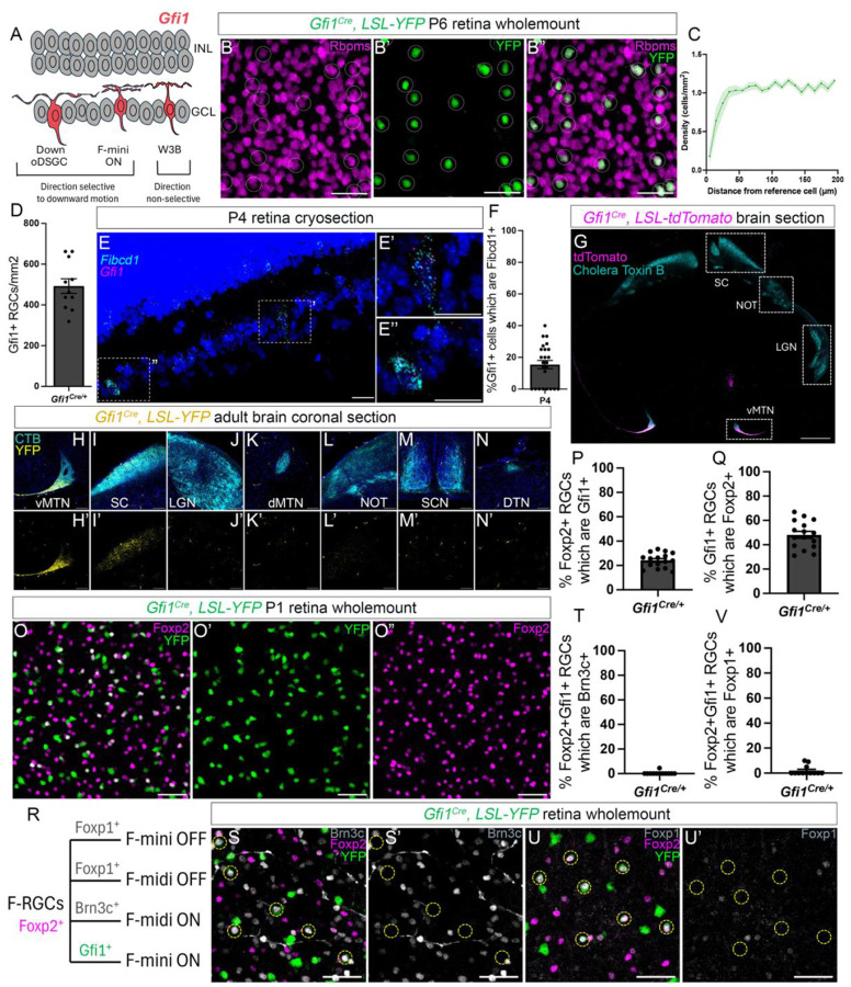
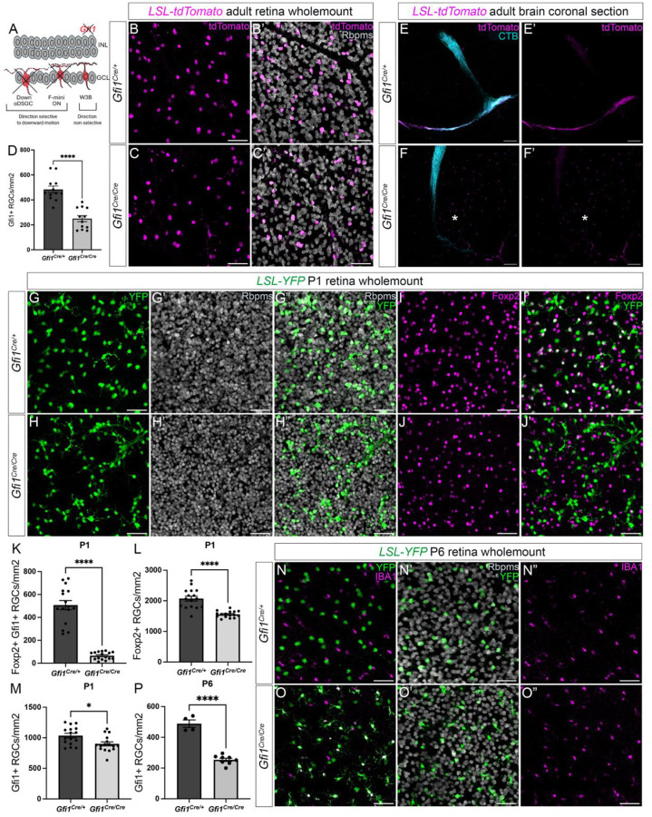
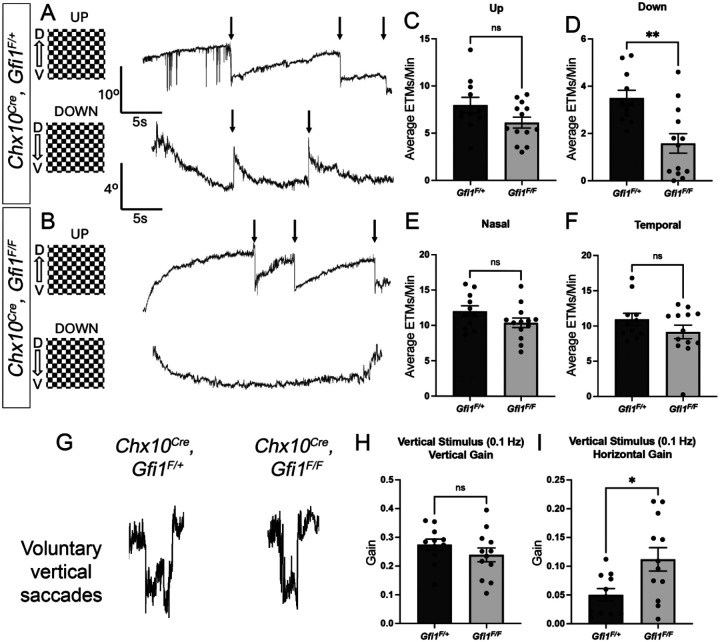
There is great diversity in retinal ganglion cells subtypes in the mouse retina, but little is known about the molecular factors required to generate this subtype diversity. Here, we identify the transcription factor Gfi1 as a conserved driver of differentiation specifically in two downward-tuned direction selective ganglion cells (DSGCs). Further, we describe a post-differentiation role for Gfi1 in regulating dendritic development of Down ON-type DSGCs crucial for their ability to detect downward motion. These results define novel temporally-segregated dual functions for Gfi1 in the development of retinal direction-selective circuits, and they provide a framework for understanding fundamental mechanisms underlying direction-selectivity in the retina.


Conflict of interest statement

Declaration of Interests The authors declare no competing interests.

Figures

Figure 1.
Figure 1.. Gfi1 is expressed in direction-selective D-oDSGC and F-mini ON RGC subsets
(A) Schematic depicting Gfi1 expression in the retina restricted to D-oDSGCs, F-mini-ONs and W3Bs. D-oDSGCs and F-mini ONs are direction-selective subsets tuned primarily to downward motion, while W3Bs are non-direction-selective. (B) Wholemount P6 Gfi1Cre, LSL-YFP retina shows YFP expression restricted to Rbpms+ RGCs. (C) Density recovery profile (DRP) of YFP+ RGCs suggests an absence of mosaicism confirming Gfi1 expression in more than one RGC subtype. (D) Density of Gfi1-expressing RGCs in the retina. (E) In situ hybridization in P4 retina cryosection shows Gfi1 expression in Fibcd1+ D-oDSGCs. (E’, E”) Detailed views of the nuclei indicated in D. (F) Quantification of the percentage of Gfi1+ RGCs co-expressing Fibcd1. (G) Coronal brain section from Gfi1Cre, LSL-tdTomato adult mouse shows strong innervation of the ventral medial terminal nucleus (MTN) by tdTomato+ RGCs. (H-N) Detailed views of all retinorecipient nuclei labeled with cholera toxin B (CTB) from Gfi1Cre, LSL-YFP adult mice confirms YFP+ RGC innervation exclusively in the ventral MTN (H, H’) and the SC (I, I’), and not in the other retinorecipient nuclei (J-N). (O) Wholemount P1 Gfi1Cre, LSL-YFP retina shows YFP expression in a subset of Foxp2+ RGCs. (P, Q) These YFP+ Foxp2+ RGCs account for ~25% of all Foxp2+ RGCs and ~50% of all Gfi1+ RGCs. (R) Combinatorial labeling can differentiate between F-RGC subsets. (S-U) Wholemount Gfi1Cre, LSL-YFP retina shows that YFP+ Foxp2+ RGCs do not express the F-RGC OFF marker Foxp1 or the F-midi ON marker Brn3c, confirming their F-mini ON identity. Data are presented as mean ± SE.
Figure 2.
Figure 2.. Gfi1 is essential for the specification of D-oDSGCs and F-mini ONs
(A) Schematic depicting a selective requirement for Gfi1 in the specification of the direction-selective D-oDSGC and F-mini-ON subsets. (B-D) Adult Gfi1Cre/Cre, LSL-tdTomato retinas show a loss of ~50% of tdTomato+ RGCs. (E-F) Gfi1Cre/Cre, LSL-tdTomato mice reveal a loss of D-oDSGCs, confirmed by the lack of ventral MTN innervation (indicated by asterisks). (G-J) P1 retinas from Gfi1Cre/Cre, LSL-YFP mice show a ~25% reduction in Foxp2+ RGCs (L), reflecting the loss of YFP+ Foxp2+ F-mini ONs (I-K), but only a small change in YFP+ RGCs (M), suggesting the presence of improperly specified RGCs. Note the excessive clumping of YFP+ RGCs (H). (N-P) By P6, the ~50% reduction in YFP+ RGCs is apparent, suggesting a loss of these improperly specified RGCs between P1 and P6. Data are presented as mean ± SE. *p < 0.05, ****p < 0.0001.
Figure 3.
Figure 3.. Gfi1 conditional mutants show selective deficits in downward motion detection
(A-F) Optokinetic reflex (OKR) results in adult Chx10Cre, Gfi1F/+ and Chx10Cre, Gfi1F/F mice in response to upward (C), downward (D), forward (E), and backward (F) continuous motion, quantified as eye-tracking movements (ETMs) per minute (arrows in A and B). Chx10Cre, Gfi1F/F mice show a selective deficit in downward OKR (B, D). (G) Chx10Cre, Gfi1F/+ and Chx10Cre, Gfi1F/F mice both performed voluntary vertical saccades. (H, I) Chx10Cre, Gfi1F/F mice show no change in vertical gain (H) but an enhanced horizontal gain (I) in response to vertical sinusoidal stimuli. Data are presented as mean ± SE. *p < 0.05, **p < 0.01.
Figure 4.
Figure 4.. Gfi1 conditional removal preserves D-oDSGC identity but disrupts function
(A) Schematic depicting retrograde injection of 555-retrobeads into the MTN to assess MTN-projecting RGC density (B, C) Chx10Cre, Gfi1F/F mice show intact ventral MTN innervation suggesting no loss of D-oDSGCs. (D-G) Wholemount retinas of Chx10Cre, Gfi1F/+ and Chx10Cre, Gfi1F/F mice following retrograde injection of 555-retrobeads into the MTN confirm no change in MTN-projecting RGC density (F) or U- and D-oDSGC couplets (G, dashed circles in D and E). (H) Schematic depicting transsynaptic viral labeling in the MTN via intravitral injections of anterograde monosynaptic tracer WGA-mCherry. (I-M) Transsynaptic labeling shows no change in WGA+ cell density within the MTN upon conditional removal of Gfi1. (N-Q) Chx10Cre, Gfi1F/+ mice demonstrate strong vertical motion-induced activation of the ventral MTN (outlined in white) estimated from Fos expression (N, O and Q) unlike Chx10Cre, Gfi1F/F mice which show no ventral MTN activation (P, Q). Levels of Fos in the ventral MTN show a strong correlation with strength of the downward OKR response (R). Data are presented as mean ± SE. **p < 0.01, ***p < 0.001.
Figure 5.
Figure 5.. D-oDSGCs in Gfi1 conditional mutants show a marked reduction in size and complexity of their dendritic arbors.
(A) Schematic of viral retrograde filling of MTN-projecting U-oDSGCs (Spig1GFP+) and D-oDSGCs (Spig1GFP−). (B-G) Spig1GFP− D-oDSGCs in Chx10Cre, Gfi1F/F mice show no changes in their dendritic fraction stratifying in either S4 (F) or S2 (G). S4 and S2 layers are identified by staining for cholinergic SACs that stratify in these layers. (H) D-oDSGCs do however display a significant reduction in their total dendritic length. (I, J) Schematic (I) and P7 wholemount retina (J) depicting viral retrograde filling of dual-labeled MTN-projecting U-oDSGCs and single-labeled MTN-projecting D-oDSGCs. (K-Q) Chx10Cre, Gfi1F/F mice show similar dendritic field areas of Spig1GFP+ U-oDSGCs (K, N, Q), but significantly smaller dendritic field areas of Spig1GFP− D-oDSGCs (L, M, O, P, Q). Many D-oDSGCs display short and poorly elaborated dendrites (O). (R-T) Dendritic tracing of D-oDSGCs reveals a sharp reduction in total dendritic length (Q), dendritic volume (R), and complexity (S). Data are presented as mean ± SE. *p < 0.05, ***p < 0.001, ****p < 0.0001.
Figure 6.
Figure 6.. Summary of Gfi1 functions in retinal direction-selective circuits
(A) Schematic representation of the dual functions of Gfi1 in (i) early specification of direction-selective D-oDSGCs and F-mini ONs, and (ii) late dendritic development of D-oDSGCs and possibly, F-mini ONs. (B) Loss of Gfi1’s early function in Gfi1 global mutants results in a specific loss of direction-selective D-oDSGCs and F-mini ONs. (C) Conditional Gfi1 mutants show a differential loss of Gfi1’s early function where late-born F-mini ONs are lost but early-born D-oDSGCs persist, revealing a late post-differentiation function of Gfi1 in regulating D-oDSGCs dendritic development and possibly their upstream synaptic connectivity. (D) Summary of the phenotypes seen in conditional Gfi1 mutants including loss of downward motion detection in OKR behavior and smaller and less branched D-oDSGC dendritic arbors.

Similar articles

References

    1. Kodama T. and du Lac S., Adaptive Acceleration of Visually Evoked Smooth Eye Movements in Mice. J Neurosci, 2016. 36(25): p. 6836–49. - PMC - PubMed
    1. Harris S.C. and Dunn F.A., Asymmetric retinal direction tuning predicts optokinetic eye movements across stimulus conditions. Elife, 2023. 12. - PMC - PubMed
    1. Vaney D.I., Sivyer B., and Taylor W.R., Direction selectivity in the retina: symmetry and asymmetry in structure and function. Nat Rev Neurosci, 2012. 13(3): p. 194–208. - PubMed
    1. Hamilton N.R., Scasny A.J., and Kolodkin A.L., Development of the vertebrate retinal direction-selective circuit. Dev Biol, 2021. 477: p. 273–283. - PMC - PubMed
    1. Dhande O.S., et al. , Genetic dissection of retinal inputs to brainstem nuclei controlling image stabilization. J Neurosci, 2013. 33(45): p. 17797–813. - PMC - PubMed

Publication types

LinkOut - more resources