[Preprint]. 2025 Jun 8:2025.06.04.657901.
doi: 10.1101/2025.06.04.657901.

A human ex vivo model of radiation-induced skin injury reveals p53-driven DNA damage signaling and recapitulates a TGFβ fibrotic response

Affiliations

A human ex vivo model of radiation-induced skin injury reveals p53-driven DNA damage signaling and recapitulates a TGFβ fibrotic response

Caroline Dodson et al. bioRxiv. .

Abstract

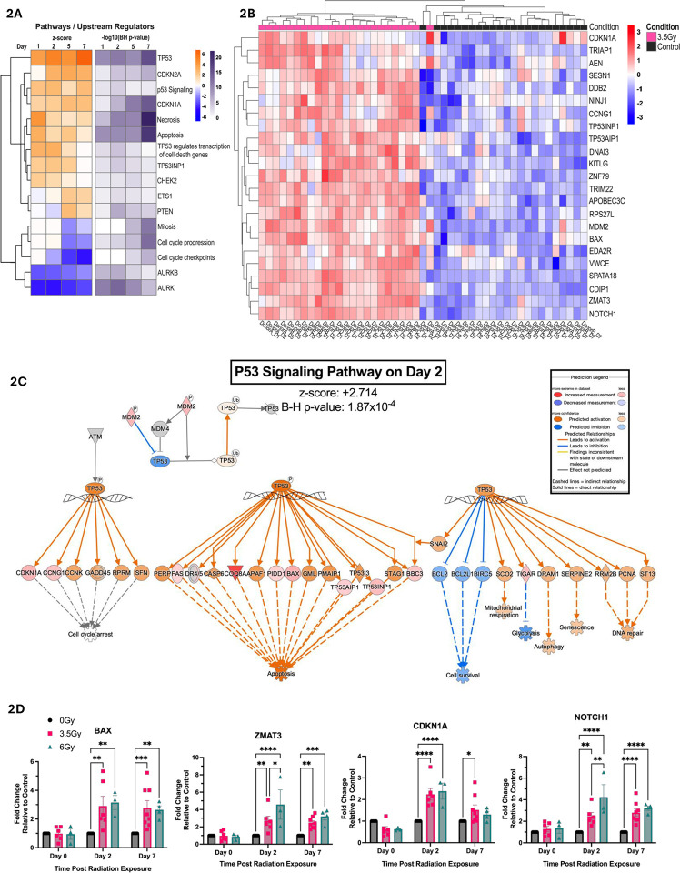
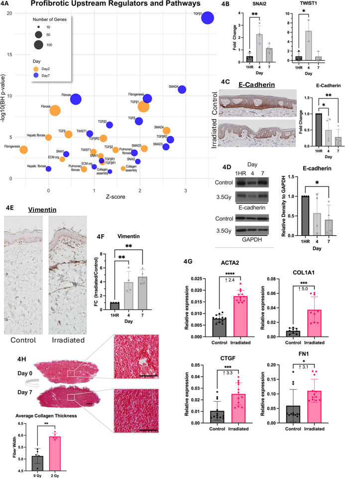
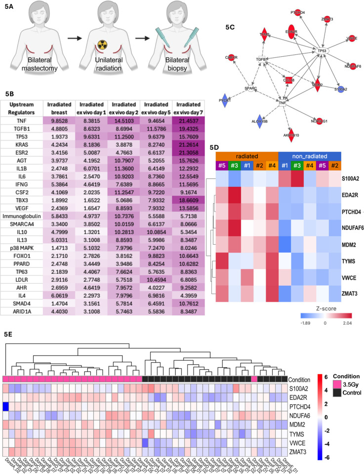
Radiation-induced skin injury is a poorly understood complication affecting cancer patients who undergo radiotherapy, with no current therapies able to prevent or halt its progression to debilitating radiation-induced skin fibrosis (RISF). Addressing the need for clinically relevant human models, this study developed and characterized a human ex vivo skin model that recapitulates the temporal molecular processes of cutaneous radiation injury, as demonstrated through bulk RNA-sequencing and tissue validation studies. Human skin explants subjected to ionizing radiation demonstrated rapid induction of DNA double-strand breaks, followed by a robust, p53-driven transcriptional program involving genes related to cell cycle arrest, apoptosis, and senescence. Over time, the irradiated skin exhibited increasing activation of pro-fibrotic pathways, notably epithelial-mesenchymal transition and TGFβ1-mediated signaling. This resulted in upregulation of classic fibrosis markers such as COL1A1, FN1, and increased collagen thickness. Importantly, regulators of the p53 axis, MDM2 and miR-34a, was observed, implicating these factors as potential therapeutic targets to modulate the balance between repair of radiation injury and pathologic fibrosis. Transcriptome analysis of irradiated and non-irradiated breast skin from post-mastectomy patients showed notable concordance of p53 and pro-fibrotic gene signatures comparable to the ex vivo model, underscoring its translational relevance. This work provides a platform for identifying early biomarkers and testing therapeutic strategies to prevent or mitigate cutaneous radiation toxicities, including RISF, beginning with elucidating the dynamic interplay between the p53-mediated DNA damage response and the onset of fibrosis following radiation. Ultimately, this work aims to improve long-term skin health and quality of life for cancer patients.


Conflict of interest statement

COMPETING INTERESTS: None

Figures

Fig. 1.
Fig. 1.. Establishment of a human ex vivo skin model and validation of early DNA damage responses to study radiation-induced injury.
(A) Workflow schematic of the human ex vivo model used in this study (B) Donor demographics (ID, age and sex) and irradiation dose. Donors A-F, marked with an asterisk, were used for bulk RNA sequencing. (C) Representative immunofluorescence images at 1-hour post-irradiation showing phospho-γH2AX (red) as a marker of double-strand DNA breaks, DAPI (blue) nuclear counterstain, and merged images from irradiated (3.5 Gy) and control (non-irradiated) samples. (D) Quantification of phospho-γH2AX staining from (C), based on counts from three independent, blinded observers. Data represents N=3 donors; symbols correspond to individual donors; mean ± SD. Statistical significance was assessed using a paired t-test. (E) Ingenuity Pathway Analysis (IPA) of gene expression data in skin immediately post-irradiation, highlighting cell-survival-related genes and pathways. Differential expression was performed using edgeR on bulk RNA-seq data filtered for protein-coding genes. IPA Core Analysis was filtered to genes with p ≤ 0.05 and log2FC ≥ ± 0.5. Gene color reflects z-score; numerical values indicate Benjamini-Hochberg-corrected −log10 (p-value). Asterisks indicate statistical significance (* ρ < 0.05, ** ρ < 0.01).
Fig. 2.
Fig. 2.. p53-Mediated DNA damage response in irradiated ex vivo skin.
(A) Ingenuity Pathway Analysis (IPA) of predicted upstream regulators and biological pathways associated with DNA damage responses across days 1, 2, 5 and 7 post-irradiation. The first heatmap is colored by IPA z-score (predicted activation state), while the second shows Benjamini-Hochberg-corrected –log10 p-values (e.g., 1.3 corresponds to p ≤0.05). Differentially expressed genes were filtered for protein-coding, p ≤ 0.05, and log2FC ≥ ±0.5. Z-score heatmap rows were clustered by decreasing row average, with p-value heatmap rows ordered to match. (B) Heatmap of TP53-regulated genes from RNA-seq data, showing row-scaled log2-transformed CPM values. Red indicates increased expression; blue indicates decreased expression. Columns represent individual samples labeled by donor and time point (e.g., DonorA_D1, DonorB_D7). Annotation bars indicate condition above the heatmap (pink = 3.5Gy, black = control). (C) IPA-annotated “p53 signaling pathway” overlaid with RNA-seq gene expression changes at day 2 post-irradiation. Genes are colored by predicted activation state (orange = activated, blue = inhibited). (D) Quantitative PCR (qPCR) of selected p53 target genes in human ex vivo skin at days 0, 2 and 7 following 0 Gy (black, N=6), 3.5 Gy (pink, N=6) or 6 Gy (teal, N=3–4) radiation. Data were analyzed using the ΔΔCt method, normalized to GAPDH, and presented as fold change relative to matched 0 Gy controls for each donor and time point; mean ± SD. Statistical significance was assessed using a mixed-effects model with Tukey’s test for multiple comparisons. Asterisks indicate statistical significance (* ρ < 0.05, ** ρ < 0.01, *** ρ < 0.001, **** ρ <0.0001). All heatmaps were generated in RStudio using the pheatmap package.
Fig. 3.
Fig. 3.. Regulators of p53-mediated DNA damage response.
(A) Quantitative PCR (qPCR) of MDM2 expression in human ex vivo skin with 0 Gy (black, N=6), 3.5 Gy (pink, N=6) and 6 Gy (teal, N=3–4) at days 0, 2 and 7 post-irradiation. Expression was normalized to GAPDH, analyzed using the ΔΔCt method and presented as fold change relative to the 0 Gy control at each time point. (B) Representative western blot showing MDM2 protein expression at day 7 in control and 3.5 Gy samples. GAPDH served as the loading control. (C) Quantification of MDM2 protein levels (B). Band intensities were extracted using ImageJ, and signals from multiple MDM2 isoforms (post-translationally modified) were averaged for each sample. Values were normalized to GAPDH. Each donor is represented by a unique symbol (N=3). (D) Representative immunofluorescence staining of phospho-MDM2 (Ser395) at day 1 post-irradiation, showing cytoplasmic localization in control samples and increased nuclear localization in irradiated samples (3.5 Gy and 6 Gy). (E) Quantification of phospho-MDM2 nuclear localization from (D), expressed as the percentage of cells with nuclear localization. Data were collected by three independent, blinded observers and analyzed using two-way ANOVA with Tukey’s multiple comparisons test (N=3 donors; symbols correspond to individual donors. Donor shown in panel D is marked with a circle). (F) RNA-seq trajectory plot showing log2 fold change in MIR34AHG expression over time, scaled to the earliest time point. (G) qPCR analysis of mature miR-34a-5p expression using cDNA enriched for small RNAs. Expression was normalized to SNORD48 and analyzed using the ΔΔCt method. Fold change was calculated relative to the 0 Gy control at each time point. (H) IPA generated network of genes regulated by miR-34 at day 7 post-irradiation. Functional annotations highlight associations with apoptosis, cell cycle, fibrosis, EMT and DNA repair. Asterisks indicate statistical significance (* ρ<0.05, ** ρ<0.01, *** ρ<0.001, **** ρ<0.0001).
Fig. 4.
Fig. 4.. Induction of epithelial-mesenchymal transition (EMT) and pro-fibrotic genes in irradiated ex vivo skin.
(A) Bubble plot showing enrichment of pro-fibrotic pathways, biological processes and upstream regulators at day 2 (yellow) and day 7 (blue) post-irradiation. The x-axis represents the predicted activation z-score, y-axis shows the Benjamini-Hochberg-corrected –log10 p-value, and bubble size reflects the number of overlapping genes. Data derived from IPA and plotted using ggplot2. (B) Quantitative PCR (qPCR) of EMT-inducing transcription in control (0 Gy) and 3.5 Gy-irradiated skin at hour 1, day 4 and day 7 post-irradiation. Expression was normalized to 18S and presented as fold change relative to control at each time point. (C) Representative IHC of E-cadherin in control and irradiated skin at day 7 post-irradiation. Quantification shown as fold change relative to control at each time point (mean ± SD; N=4). (D) Western blot of E-cadherin protein expression at 1 hour, day 4 and day 7 post-irradiation in control and 3.5 Gy samples. GAPDH was used as the loading control. Quantification of E-cadherin western blot shown as fold change relative to control at each time point (mean ± SD; N=3). (E) Representative immunohistochemistry (IHC) of vimentin in control and irradiated skin at day 7 post-irradiation. (F) Quantification of vimentin IHC over time, shown as fold change relative to control (N=4). (G) qPCR of pro-fibrotic genes in control and 3.5 Gy irradiated skin at day 7 post-irradiation. Expression was normalized to GAPDH and shown as relative expression (N=5). (H) Picrosirius red staining of 2 Gy-irradiated skin at day 0 and day 7 post-irradiation. Quantification of average collagen fiber thickness. Asterisks indicate statistical significance (* ρ < 0.05, ** ρ < 0.01, *** ρ < 0.001, **** ρ <0.0001). Statistical analyses: One-way ANOVA with Tukey’s correction for (B), (C), (D) and (F); two-tailed paired t-tests for (G) and (H).
Fig. 5.
Fig. 5.. Correlation of ex vivo human skin radiation response with irradiated breast skin from patients post-radiotherapy.
(A) Skin biopsies of the irradiated breast skin and non-irradiated skin of the contralateral breast were obtained from N=5 post-mastectomy patients at the time of surgical reconstruction. Raw bulk RNAseq profiles (GSE278183) were re-processed and re-analyzed using IPA. (B) Top 25 enriched upstream regulators common to irradiated breast skin and ex vivo irradiated skin across days 1, 2, 5, 7, ranked by sum of Benjamini-Hochberg (BH) enrichment p-values across all time points. Matrix values represent –log10(BH p-value). Colored by conditional formatting (0=white, max value=purple). (C) Network of the 15 genes overlapping between breast and at least 2 timepoints. Eight are downstream of TP53 and 5 connect to TGFβ1/SPARC (role in ECM remodeling). Red indicates commonly upregulated; blue is commonly downregulated across datasets. (D) Heatmap of 8 commonly regulated TP53 genes across profiles from non-irradiated and irradiated breast skin (patients numbered #1-#5). (E) Heatmap of the same TP53 genes from panel (D) in human ex vivo skin. Data were log2-transformed counts per million (CPM) values. Red indicates increased expression; blue indicates decreased expression. Annotation bars indicate the condition above the heatmap (pink = 3.5 Gy, black = control). Heatmap was generated in Rstudio using the pheatmap package.

References

    1. Baskar R., Lee K. A., Yeo R., Yeoh K. W., Cancer and radiation therapy: current advances and future directions. Int J Med Sci 9, 193–199 (2012). - PMC - PubMed
    1. Borrelli M. R. et al. , Radiation-Induced Skin Fibrosis: Pathogenesis, Current Treatment Options, and Emerging Therapeutics. Ann Plast Surg 83, S59–S64 (2019). - PMC - PubMed
    1. Bentzen S. M., Preventing or reducing late side effects of radiation therapy: radiobiology meets molecular pathology. Nat Rev Cancer 6, 702–713 (2006). - PubMed
    1. Ryan J. L., Ionizing radiation: the good, the bad, and the ugly. J Invest Dermatol 132, 985–993 (2012). - PMC - PubMed
    1. Stone H. B., Coleman C. N., Anscher M. S., McBride W. H., Effects of radiation on normal tissue: consequences and mechanisms. Lancet Oncol 4, 529–536 (2003). - PubMed

Publication types