[Preprint]. 2025 Jun 4:2025.05.29.25328544.
doi: 10.1101/2025.05.29.25328544.

Mosaic chromosomal alterations in blood are associated with an increased risk of Alzheimer's disease

Affiliations

Mosaic chromosomal alterations in blood are associated with an increased risk of Alzheimer's disease

Tatsuhiko Naito et al. medRxiv. .

Abstract

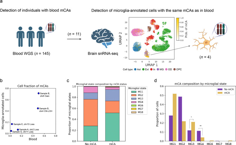
Mosaic chromosomal alterations (mCAs) in blood, a form of clonal hematopoiesis, have been linked to various diseases, but their role in Alzheimer's disease (AD) remains unclear. We analyzed blood whole-genome sequencing (WGS) data from 24,049 individuals in the Alzheimer's Disease Sequencing Project and found that autosomal mCAs were significantly associated with increased AD risk (odds ratio = 1.27; P = 1.3 × 10⁻⁵). This association varied by ancestry, mCA subtype, APOE ε4 allele status, and chromosomal location. Using matched blood WGS and brain single-nucleus RNA-seq data, we identified microglia-annotated cells in the brain carrying the same mCAs found in blood. These findings suggest that blood mCAs may contribute to AD pathogenesis, potentially through infiltration into the brain and influencing local immune response.


Conflict of interest statement

Competing interests The authors declare no conflicts of interest for this study. T.R. served as a scientific advisor for Merck and serves as a consultant for Curie.Bio.

Figures

Figure 1.
Figure 1.. The association between autosomal mCAs and AD.
a. The prevalence of mCAs by age. The solid and dashed lines represent AD cases and controls, respectively. Colors indicate mCA types. b. Forest plots showing the odds ratio (ORs) of mCAs for AD in each ancestry group, with results presented by mCA type. c. Forest plot showing the ORs of mCAs for AD in individuals with and without the APOE ε4 allele. d. Forest plot showing the ORs for the presence of 1, 2, or ≥3 mCAs for AD in individuals without the APOE ε4 allele. e. Heatmap showing the ORs of mCAs on each chromosome for AD. f. The prevalence of CHIP by age. The solid and dashed lines represent AD cases and controls, respectively. g. Forest plot showing the ORs of CHIP for AD in each ancestry group. In all plots, the error bars represent 95% confidence intervals. *, **, and *** represent P-value in association test < 0.05, 0.01, and 0.001, respectively. ALL, all ancestries; AFR, African American; AMR, Admixed American; EUR, European; SAS, South Asian; AD, Alzheimer’s disease; OR, odds ratio.
Figure 2.
Figure 2.. Detection of the same mCA as identified in blood from brain snRNA-seq data.
a. Using individuals with paired blood WGS and brain snRNA-seq data, we first identified those carrying blood mCAs, and then detected cells harboring the same mCAs, which were markedly enriched within the microglia-annotated population. The central uniform manifold approximation and projections (UMAP) plot shows the probability of harboring mCAs overlaid on cell type annotations in samples where mCAs were significantly enriched in microglia-annotated cells. b. Scatter plot showing the cell fractions of mCAs in blood and microglia-annotated cells. c. Bar plot showing the proportion of microglial states by mCA status. d. Bar plot showing the proportion of cell with and without mCA across different microglial states. Some illustrations were made with the aid of BioRender. Ast, astrocytes; Ext, excitatory neurons; In, inhibitory neurons; MG, microglia; OD, oligodendroglia; OPC, oligodendrocyte progenitor cells.

References

    1. Jaiswal S. & Ebert B. L. Clonal hematopoiesis in human aging and disease. Science 366, (2019). - PMC - PubMed
    1. Brown D. W. et al. Shared and distinct genetic etiologies for different types of clonal hematopoiesis. Nat. Commun. 14, 5536 (2023). - PMC - PubMed
    1. Xie M. et al. Age-related mutations associated with clonal hematopoietic expansion and malignancies. Nat. Med. 20, 1472–1478 (2014). - PMC - PubMed
    1. Jaiswal S. et al. Clonal Hematopoiesis and Risk of Atherosclerotic Cardiovascular Disease. N. Engl. J. Med. 377, 111–121 (2017). - PMC - PubMed
    1. Miller P. G. et al. Association of clonal hematopoiesis with chronic obstructive pulmonary disease. Blood 139, 357–368 (2022). - PMC - PubMed

Publication types

LinkOut - more resources