[Preprint]. 2025 Jun 6:rs.3.rs-6710731.
doi: 10.21203/rs.3.rs-6710731/v1.

Depth-Variant Deconvolution Applied to Widefield Microscopy for Rapid Large-Volume Tissue Imaging

Affiliations

Depth-Variant Deconvolution Applied to Widefield Microscopy for Rapid Large-Volume Tissue Imaging

Daniel D Lee et al. Res Sq. .

Abstract

Innovations in 3D tissue imaging have revolutionized research, but limitations stemming from lengthy protocols and equipment accessibility persist. Classical widefield microscopy is fast and accessible but often excluded from 3D imaging workflows due to its lack of optical sectioning. Here we combine tissue clearing with a depth-variant deconvolution approach customized for large-volume widefield imaging to achieve subnuclear axial resolution in tissues to a depth of 500 μm. We illustrate the utility of this method in a mouse model of ileitis and to gain a 3D perspective in thick brain slices from a mouse model of cerebral amyloid angiopathy, where we resolved large and small blood vessels, including those with amyloid deposits, attaining resolution that compared favorably to tile-scanning confocal microscopy. Finally, we sought to leverage our approach to allow for richer pathological evaluation of human kidney biopsies. Our approach produced hundreds of consecutive z-planes in five minutes of imaging for 3D visualization of winding arterioles feeding into glomeruli. This 3D perspective afforded straightforward identification of atrophic tubes in fresh kidney biopsies prepared in 2 hours to simulate the time-constrained evaluation of donor kidneys for transplant suitability. Having achieved subnuclear z-resolution in sections hundreds of microns thick, widefield microscopy coupled to robust deconvolution now emerges as an accessible and viable method to gain 3D insight in research or clinical pathological evaluations.


Conflict of interest statement

Additional Declarations: Yes there is potential Competing Interest. Some of the authors have filed a provisional patent for the ADAPT-3D clearing method used in this manuscript. Declaration of Interests: Some of the authors have filed a provisional patent for the ADAPT3D clearing method used in this manuscript.

Figures

Figure 1.
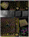
Figure 1.. Depth variant deconvolution of thick image volumes acquired with a widefield microscope achieves sub-nuclear axial resolution for 3D visualization.
(A) Standard epifluorescent microscopy build with 20X immersion lens (Carl Zeiss AG, NA 1.0, working distance of 6.4 mm) (top). Optical path for a refractive index matched tissue mounted under glass coverslip with 20X lens that is immersed in water (bottom). (B) Extended display showing raw image of nuclei in a cleared mouse brain labeled with anti-Histone-H3-ATTO488 (green) acquired on widefield microscope before computational deconvolution. (C) Nuclei in cleared brain after deconvolution with depth-variant point spread functions calculated using Huygens Software (Scientific Volume Imaging) and visualized in the axial dimension with Imaris software. (D) Extended display showing resolution of fine microglial pseudopods labeled by Cx3cr1-tdTomato (green) and nuclear staining with anti-Histone-H3-ATTO488 (magenta) captured using epifluorescent widefield microscopy with 20X (NA 1.0) lens; (E) Extended display of microglia as in (D) but captured using line-scanning confocal microscopy at a similar pixel resolution. (F) Maximum intensity projection of ~200 microns of 5XE4 leptomeninges attached to the brain surface that were stained for Aβ (red) to label CAA plaques along arterioles (smooth-muscle actin: green, CD31-positive vessels: gray), imaged using epifluorescent widefield microscopy with 5X objective lens (NA 0.16, air), and deconvolved.
Figure 2.
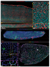
Figure 2.. Optimizing deconvolution of deep tissue widefield imaging to enable tiling for large volume imaging
(A) 3D display of single tile depicting striping edge artifacts (depicted by arrows) following deconvolution from mouse ileum immunostained with LYVE1 (magenta), smooth muscle actin (SMA, yellow), and S100A9 (cyan). (B) Extended display of same deconvolved tile as in (A) following cropping of artifacts as indicated with dashed lines and overview 3D display in (B’). (C) Extended display of the same tile in (A) when local background is subtracted before the deconvolution process without any cropping and a 3D overview (C’). (D) x-y projection of 142 microns depth of brain capillaries immunostained with anti-CD31 and anti-podocalyxin before background subtraction and (E) after subtraction of background found with a combined gaussian and minimum filter. (F) 3D projection of brain cortex following deconvolution of the whole volume as a single brick or (G) after subdividing the volume into 7 separate bricks in the Z axis for maximal retention of fine capillaries found in the raw image (D). (H) Extended display of a stitched 3x3 tile of mouse ileum after background subtraction and deconvolution with z bricks immunostained for LYVE1, SMA, and S100A9. (I) 3D display of entire overview from mouse ileum in (H). (J) x-y projection of 50 microns from ileum that depicts fine resolution of neutrophils in muscularis layer of mouse ileum from (H-I).
Figure 3.
Figure 3.. Large volume 3D widefield imaging enables comprehensive overview of CAA pathology at sub-nuclear resolution
(A) Representative x-y projection of blood vasculature (CD31 and podocalyxin, red, and Histone-H3-ATTO488, cyan) from a 15-tile acquisition of a sagittal 500-micron section of mouse brain imaged, deconvolved, and stitched. (B) Extended display of image in panel (A) illustrating the depth and resolution of fine capillaries and nuclei. (C) Tilted 3D display of 80-tile image of Aβ plaques (green) in association with vessels (CD31/podocalyxin, blue) or arterioles (SMA, red) across sagittal section of brain isolated from 7-month old 5XE4 mouse acquired using 20X immersion lens, deconvolved, and stitched. * indicates bridging vein with deposits of Aβ plaques in SMA-negative vessel (i.e., bridging vein) accompanied by a SMA-positive vessel arteriole along the leptomeninges surface. (D) Extended display depicting three-dimensional CAA of capillaries zoomed in from (C, dashed box). (E) Maximum intensity projection after deconvolution of leptomeninges on the surface of an uncleared brain hemisphere from a 9-month old CAA mouse labeled for SMA (yellow), CD31 (cyan), and Aβ (magenta) and acquired using a 5X air objective (NA 0.16). Arrow indicates a bridging vein with Aβ deposition.
Figure 4.
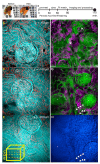
Figure 4.. Rapid optical transparency and widefield imaging facilitates 3D evaluation of human kidney biopsies for pathology in a pre-transplant scenario
(A) ~0.45 mm wedge biopsy treated with periodic acid, stained with CellBrite Orange, FAM-Hydrazide, and DAPI before refractive index matching (left) and after 30 minutes of refractive index matching (right). (B) Timeline overview to obtain optical transparency of wedge biopsy as seen in (A). (C-E) 5 μm X-Y maximum intensity projections of 3 separate glomeruli spaced at 100 μm z-intervals extracted from a single imaging volume of a kidney biopsy labeled with a Periodic Acid Schiff stain (cyan) and nuclei (red). Lower box schematic (yellow) of image volume from which digital sections were extracted at different depths. (F) Digital section from 3D image of biopsy constrained to timing shown in (B) depicting intact glomeruli and its neighboring proximal tubules, some of which contain atrophy (arrows). (G) 3 intact glomeruli (*), along with a possible arteriosclerotic vessel (arrowheads) found 100 microns of Z-depth below the digital section in (F) and its accompanying DAPI nuclear staining (H).

References

    1. Matsumoto K, Mitani TT, Horiguchi SA, Kaneshiro J, Murakami TC, Mano T, Fujishima H, Konno A, Watanabe TM, Hirai H, Ueda HR: Advanced CUBIC tissue clearing for whole-organ cell profiling. Nat Protoc 2019, 14:3506–37. - PubMed
    1. Erturk A, Becker K, Jahrling N, Mauch CP, Hojer CD, Egen JG, Hellal F, Bradke F, Sheng M, Dodt HU: Three-dimensional imaging of solvent-cleared organs using 3DISCO. Nat Protoc 2012, 7:1983–95. - PubMed
    1. Hsu CW, Cerda J 3rd, Kirk JM, Turner WD, Rasmussen TL, Flores Suarez CP, Dickinson ME, Wythe JD: EZ Clear for simple, rapid, and robust mouse whole organ clearing. Elife 2022, 11. - PMC - PubMed
    1. Lee DD, Davis DL, Smyth LCD, Telfer KA, Ravindran R, Czepielewski RS, Huckstep CG, Kurashima K, Jain AK, Kipnis J, Zinselmeyer BH, Randolph GJ: ADAPT-3D: Accelerated Deep Adaptable Processing of Tissue for 3-Dimensional Fluorescence Tissue Imaging for Research and Clinical Settings. Research Square (preprint server) 2025. - PMC - PubMed
    1. Shaw PJ: Comparison of Widefield/Deconvolution and Confocal Microscopy for Three-Dimensional Imaging. Handbook of Biological Confocal Microscopy. Edited by Pawley JB. 3rd ed. New York: SpringerScience+Business Media, 2006. pp. 453–67.

Publication types

LinkOut - more resources