Case Reports
. 2024 Jun;24(2):182-186.
doi: 10.7704/kjhugr.2024.0014. Epub 2024 Jun 10.

A Rare Case of Signet Ring Cell Carcinoma Arising on Duodenal Brunner's Gland Hyperplasia Successfully Treated Via Endoscopic Resection

Affiliations
Case Reports

A Rare Case of Signet Ring Cell Carcinoma Arising on Duodenal Brunner's Gland Hyperplasia Successfully Treated Via Endoscopic Resection

Hae Rin Lee et al. Korean J Helicobacter Up Gastrointest Res. 2024 Jun.

Abstract

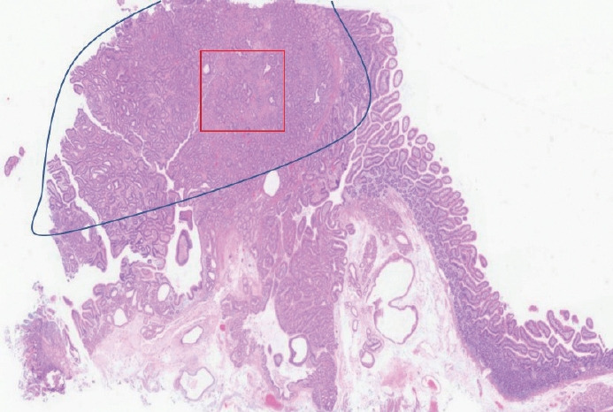
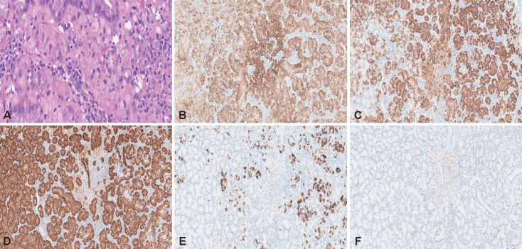
Signet-ring cell carcinoma (SRCC) is a rare tumor that most commonly occurs in the stomach. Duodenal SRCCs are extremely uncommon and account for approximately 1% of duodenal adenocarcinomas. Although Brunner's gland hyperplasia (BGH) is a benign duodenal condition, studies have reported several cases of adenocarcinoma originating in an area of BGH. We report a rare case of early-stage SRCC originating in an area of BGH that was successfully treated using endoscopic mucosal resection. Based on the mucin phenotype observed in this case, it is reasonable to conclude that SRCC originated from gastric metaplasia in the area of BGH. Although BGH is a benign condition, careful evaluation is warranted for early detection of combined neoplasms.

Keywords: Brunner’s gland; Carcinoma, Signet ring cell; Duodenum; Endoscopic mucosal resection.


Conflict of interest statement

Conflicts of Interest The authors have no financial conflicts of interest.

Figures

Fig. 1.
Fig. 1.
Endoscopic and endosonographic findings. A and B: EGD showed a 13 mm-sized subepithelial lesion, located in the second portion of the duodenum, showing a linear erosion with locally reddish irregular mucosa on the surface. C and D: Narrow-band imaging showed abnormally distorted mucosal pattern on the surface (inside the yellow line). E: EUS revealed a mass with heterogeneous echogenicity with small anechoic areas mainly located in the submucosa in favor of typical features of BGH. F: SRCC seemed to be confined to mucosa layer (inside the yellow arrow). EGD, esophagogastroduodenoscopy; EUS, endoscopic ultrasonography; BGH, Brunner’s gland hyperplasia; SRCC, signet ring cell carcinoma.
Fig. 2.
Fig. 2.
Endoscopic mucosal resection. A: Submucosal injection was done to lift the lesion. B and C: The tumor was totally resected using an endoscopic snare. D: Endoclips were placed to close the defect after EMR. E and F: Gross appearance of the resected specimen showed a 13 mm-sized subepithelial tumor, with a reddish irregular mucosal surface showing abnormal vasculature via near-focus imaging. EMR, endoscopic mucosal resection.
Fig. 3.
Fig. 3.
Gastric foveolar metaplasia overlying BGH (blue line) is identified. Signet ring cell carcinoma (red square) is found in the center (H&E; magnification, ×1.25). BGH, Brunner’s gland hyperplasia; H&E, hematoxylin and eosin.
Fig. 4.
Fig. 4.
Histopathological findings. A: Tumor cells show signet ring cell features, which are characterized by abundant intracytoplasmic mucin with an eccentrically placed nucleus (H&E; magnification, ×400). On immunohistochemical findings, tumor cells were positive for panCK (B), MUC5AC (C) and weakly positive for MUC6 (D), but negative for MUC2 (E) and CD10 (F) (magnification, ×100). H&E, hematoxylin and eosin; panCK, pan Cytokeratin; MUC5AC, mucin 5AC; MUC6, mucin 6; MUC2, mucin 2.

References

    1. Mochizuki K, Kondo T, Tahara I, et al. Signet ring cell carcinoma of the non-ampullary duodenum: a case report. Pathol Res Pract. 2015;211:801–804. - PubMed
    1. Ye N, Bao X, Zhao X, Wang B. Signet-ring cell carcinoma of the duodenal bulb presenting with gastrointestinal hemorrhage: a case report and literature review. BMC Gastroenterol. 2022;22:226. - PMC - PubMed
    1. Lee KJ, Park B, Kim HM. Endoscopic ultrasonography findings for Brunner’s gland hamartoma in the duodenum. Clin Endosc. 2022;55:305–309. - PMC - PubMed
    1. Iwamuro M, Kobayashi S, Ohara N, Kawano S, Kawahara Y, Okada H. Adenocarcinoma in situ arising from Brunner’s gland treated by endoscopic mucosal resection. Case Rep Gastrointest Med. 2017;2017:7916976. - PMC - PubMed
    1. Hizawa K, Iwai K, Esaki M, et al. Endosonographic features of Brunner’s gland hamartomas which were subsequently resected endoscopically. Endoscopy. 2002;34:956–958. - PubMed

Publication types

LinkOut - more resources