. 2025 May 28:16:1573492.
doi: 10.3389/fimmu.2025.1573492. eCollection 2025.

PD-1 is conserved from sharks to humans: new insights into PD-1, PD-L1, PD-L2, and SHP-2 evolution

Affiliations

PD-1 is conserved from sharks to humans: new insights into PD-1, PD-L1, PD-L2, and SHP-2 evolution

Ryohei Kondo et al. Front Immunol. .

Abstract

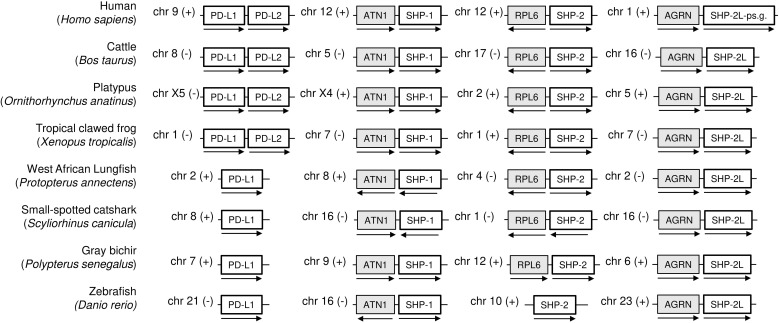
Programmed cell death protein 1 (PD-1) is an immune checkpoint molecule until recently believed to exist only in tetrapod species. However, together with a very recent study dedicated to the CD28/CTLA4 molecule family, this study-using database information-identifies the PD-1 gene in both bony and cartilaginous fish, while being the first to present a detailed molecular analysis of the evolution of PD-1 and its ligands. Conserved sequence motifs imply an ancient origin of PD-1's binding modes to its extracellular ligand PD-L1 and its intracellular ligand Src homology region 2 domain-containing phosphatase-2 (SHP-2), and also of its N116 glycosylation motif-a less well known PD-1 feature-important for binding galectins. The PD-1 cytoplasmic tail binds SHP-2 by two motifs, defined as an immunoreceptor tyrosine-based inhibitory motif (ITIM) and immunoreceptor tyrosine-based switch motif (ITSM), but sequence conservation patterns show that these definitions warrant a discussion. As in mammals, PD-1 transcripts in fish could be found co-expressed with markers of regulatory and exhausted T cells, suggesting a similar immune checkpoint function. Agreeing with previous reports, the PD-L1/PD-L2 gene duplication was only found in tetrapod species, while we newly discovered that features that consistently distinguish the two molecules are PD-L2 IgC domain motifs. Among PD-L1 (the name given to the single PD-L ancestral molecule) of many ray-finned fish, conservation of a very long cytoplasmic tail motif supports previous claims that PD-L1 cytoplasmic tails may have a function. Surprisingly, we found a gene similar to SHP-2-that we named SHP-2-like (SHP-2L)-to be conserved from sharks to mammals, although lost or inactivated in higher primates and rodents. SHP-2L is expected to bind PD-1 similar to SHP-2. This comparative analysis of PD-1 and its interacting molecules across jawed vertebrates highlights conserved immune checkpoint features while revealing new insights and lineage-specific adaptations.

Keywords: PD-1; PD-L1; PD-L2; SHP-1; SHP-2; SHP-2L; evolution; fish.


Conflict of interest statement

The authors declare that the research was conducted in the absence of any commercial or financial relationships that could be construed as a potential conflict of interest. The author(s) declared that they were an editorial board member of Frontiers, at the time of submission. This had no impact on the peer review process and the final decision

Figures

Figure 1
Figure 1
The genomic locations of PD-1 genes from shark to human, compared in seven representative species. Selected genes and their orthologues in other species that in human are linked to PD-1 on chromosome 2 are indicated as green boxes: neuronal guanine nucleotide exchange factor (NGEF); autophagy related 16 like 1 (ATG16L1); diacylglycerol kinase delta (DGKD); sushi, nidogen and EGF like domains 1 (SNED1); mitochondrial transcription termination factor 4 (MTERF4); and BCL2 family apoptosis regulator (BOK). Relevant genes located on human chromosome 3 are indicated as blue boxes: TSC22 domain family member 2 (TSC22D2); dynein light chain Tctex-type 2B (DYNLT2B); eph receptor B3b (EPHB3B); SRY-box transcription factor 2 (SOX2); and phosphate cytidylyltransferase 1A (PCYT1A). The arrows indicate the gene direction. Genes are not necessarily neighbors, and the locations on the chromosome are indicated in Mb. chr, chromosome; M, Mb; (+), forward relationship (–); reverse relationship.
Figure 2
Figure 2
Alignment of deduced PD-1 amino acid sequences in representative species of Sarcopterygii (tetrapods and lobe-finned fishes), Condrichthyes (cartilaginous fish), and Actinopterygii (ray-finned fish). The (predicted) leader sequences are shown in Italic font and gray shading. In the IgSF domain, colored shading indicates: yellow, the most characteristic residues of IgSF domains; gray, other characteristic IgSF V category residues; cyan, residues more typical of PD-1 from shark to human. In the cytoplasmic tail, colored shading indicates: yellow, residues matching ITIM and ITSM definitions (S/I/V/L)xYxx(I/V/L) and TxYxx(V/I), respectively, except for the threonine at position -2 in the ITSM motif which is colored green; cyan, other residues of the (I/V)(D/E)Y(A/G)(E/V)L(D/E)F and (T/V)EYATIx(F/Y) motifs. The sequences and their sources are shown in Supplementary File 1B . Residue numbering above the alignment follows the human PD-1 protein. The numbers between brackets refer to introns and to their phases at the indicated position (0) or in the preceding codon (1,2) in the corresponding genomic sequences. Cysteines are in purple and, based on Hopp and Woods, 1981 (82): red font is used for basic residues, blue for acidic residues, and of the other residues (green and orange) the more hydrophilic ones are in green. Underlining of human and mouse PD-1 strecthes in the ectodomain indicates β-strands (following PDB accessions 3RRQ and 1NPU), and the indication of the human transmembrane region follows UniProt accession Q15116. The species are: human (Homo sapiens), mouse (Mus musculus), cattle (Bos taurus), platypus (Ornithorhynchus anatinus), chicken (Gallus gallus), goose (swan goose; Anser cygnoides domesticus), lizard (green anole lizard; Anolis carolinensis), turtle (green sea turtle; Chelonia mydas), frog (tropical clawed frog; Xenopus tropicalis), lungfish (West African Lungfish (Protopterus annectens), shark (small-spotted catshark; Scyliorhinus canicula), skate (thorny skate; Amblyraja radiata), bichir (gray bichir (Polypterus senegalus), reedfish (reedfish; Erpetoichthys calabaricus), sturgeon (sterlet sturgeon; Acipenser ruthenus), paddlefish (Mississippi paddlefish; Polyodon spathula), gar (spotted gar; Lepisosteus oculatus), bonytongue (Asian bonytongue; Scleropages formosus), tarpon (Megalops atlanticus), weatherfish (oriental weatherfish; Misgurnus anguillicaudatus), zebrafish (Danio rerio), salmon (Atlantic salmon; Salmo salar), perch (Barramundi perch; Lates calcarifer), medaka (Oryzias latipes), mummichog (Fundulus heteroclitus).
Figure 3
Figure 3
Conservation of PD-1 and PDL-1 interaction between fish and mammals. The figures show superimpositions of the (membrane-distant) IgSF domains of human PD-1 bound to human PD-L1 (PDB accession 4ZQK) and a MultiFOLD predicted interaction structure of the corresponding domains in tarpon PD-1 and PD-L1. Depictions are in cartoon format, except for the highlighted residues that are also in sticks format. The conserved intermolecular hydrogen bonds of PD-1 Y68 with PD-L1 D122 and of PD-1 K78 and PD-L1 F19 are indicated by dashed lines. (A) Superimposition guided by the compared complex structures. The figure shows that also in tarpon the PD-1 domain is predicted to have a typical IgSF structure with conserved positions of the IgSF-typical residues G47, (I/V)110, D117, G119, Y121, and L142. (B) A superimposition guided by the membrane-distant PD-L1 IgSF domain shows how well conserved this domain is. (C) Similar as B, but highlighting the conserved intermolecular hydrogen bonds.
Figure 4
Figure 4
Schematic depiction of the genomic locations of PD-L1, SHP-1, SHP-2, and SHP-2L genes. For the same model species as shown in Figure 1 , this figure shows that (i) a second PD-L gene was only found in tetrapods, (ii) SHP-1, SHP-2, and SHP-2L are located on different chromosomes, showing regional conservation between species, and (iii) human SHP-2L is only represented by pseudogene fragments. Genes depicted on the same chromosome are not necessarily neighboring genes. ATN1, atrophin 1; RPL6, ribosomal protein L6; AGRN, agrin.
Figure 5
Figure 5
Structural position of PD-L2 residues that distinguish PD-L2 from PD-L1 throughout tetrapods. PD-L2 residues L150, G172, N189, and S191 are highly conserved among, and in tetrapods specific for, PD-L2 (C atoms in yellow). The L150 sidechain forms a complex with aromatic groups of residues at positions 166 and 174 (C atoms in cyan). Highlighted resides are shown in spheres format and element coloring (red for O, blue for N). The depicted structure shows mouse PD-1 and PD-L2 ectodomains (PDB accession 3BP5).
Figure 6
Figure 6
Impressive conservation of long PD-L1 cytoplasmic tail extensions in many ray-finned fish. This figure shows an alignment of the exon-8-encoded C-termini of the nine, out of 13, representative ray-finned fish PD-L1 sequences (see Supplementary file 2A ) in which such extension could be found. Highly conserved residues are highlighted by yellow (when identical or highly similar) or gray (when similar) shading. Font coloring of residues is as in Figure 2 .
Figure 7
Figure 7
Expression patterns of immunosuppressive genes in salmon and mouse spleens. (A) UMAP visualization of gene expression patterns for T cell markers and immunosuppressive genes across spleen cells. The color gradient indicates the normalized expression level. (B) Gene ranking based on co-expression correlation with PD-1. Genes that show the highest correlation with PD-1 (top 100) in both mouse and salmon are labeled with their ranking in parentheses. Gene identities are explained in Supplementary File 5 .
Figure 8
Figure 8
Proposed PD-1/SHP-2 interaction modes. This figure is reused from Patsoukis et al., Interaction of SHP-2 SH2 domains with PD-1 ITSM induces PD-1 dimerization and SHP-2 activation, Communications Biology, 2020a (60), under a CC BY 4.0 license. This figure was also used in Patsoukis et al., 2020b (61), together with the following explanation: (A) Two-step binding model, according to which SHP-2 C-SH2 binds to PD-1 pY-ITSM with strong affinity, recruiting PD-1 to SHP-2, while PD-1 pY-ITIM binds to N-SH2, displacing it from the PTP site [the Protein Tyrosine Phosphatase domain] to activate the phosphatase. (B) Dimerization model, according to which SHP-2 bridges two pY-ITSM residues on two PD-1 molecules via its N-SH2 and C-SH2 domains forming a PD-1:PD-1 dimer and inducing SHP-2 activation.
Figure 9
Figure 9
Structures of SHP-2 domains bound to pY peptides derived from the PD-1 cytoplasmic tail motifs. The structures are based on human molecules and were determined by Marasco et al., 2020 (15). (A) SHP-2 N-SH2 domain bound to a pY peptide derived from the PD-1 ITIM region; the structure was determined using X-ray crystallography (PDB accession 6ROY). (B) SHP-2 C-SH2 domain bound to a pY peptide derived from the PD-1 ITSM region; the structure was determined using NMR spectroscopy (PDB accession 6R5G). The depicted SHP-2 domains are from the WFH residues until the PLNC or PLNT residues (see Supplementary File 3A ), and are presented in semi-transparent surface style and element coloring (white for C, red for O, blue for N)—except for SHP-2 residues I54 and V170, which are fully in green and contribute to the specificity pockets. Black font descriptions indicate the PD-1 residues and their positions relative to the pY residues. Dashed yellow lines indicate intermolecular polar contacts, and these include interactions between the phosphate groups of the respective pY residues with R32 in N-SH2 or R138 in C-SH2, and one between ITSM-T(pY+2) and C-SH2-E204 (respective SHP-2 residues are not highlighted).

Similar articles

References

    1. Ishida Y, Agata Y, Shibahara K, Honjo T. Induced expression of PD-1, a novel member of the immunoglobulin gene superfamily, upon programmed cell death. EMBO J. (1992) 11:3887–95. doi: 10.1002/j.1460-2075 - DOI - PMC - PubMed
    1. Agata Y, Kawasaki A, Nishimura H, Ishida Y, Tsubata T, Yagita H, et al. Expression of the PD-1 antigen on the surface of stimulated mouse T and B lymphocytes. Int Immunol. (1996) 8:765–72. doi: 10.1093/intimm/8.5.765 - DOI - PubMed
    1. Latchman Y, Wood CR, Chernova T, Chaudhary D, Borde M, Chernova I, et al. PD-L2 is a second ligand for PD-1 and inhibits T cell activation. Nat Immunol. (2001) 2:261–8. doi: 10.1038/85330 - DOI - PubMed
    1. Cai L, Li Y, Tan J, Xu L, Li Y. Targeting LAG-3, TIM-3, and TIGIT for cancer immunotherapy. J Hematol Oncol. (2023) 16:101. doi: 10.1186/s13045-023-01499-1 - DOI - PMC - PubMed
    1. Twomey JD, Zhang B. Cancer immunotherapy update: FDA-approved checkpoint inhibitors and companion diagnostics. AAPS J. (2021) 23:39. doi: 10.1208/s12248-021-00574-0 - DOI - PMC - PubMed

MeSH terms

Substances

LinkOut - more resources