. 2025 Jun 17;122(24):e2426528122.
doi: 10.1073/pnas.2426528122. Epub 2025 Jun 12.

SARS-CoV-2 nsp15 enhances viral virulence by subverting host antiviral defenses

Affiliations

SARS-CoV-2 nsp15 enhances viral virulence by subverting host antiviral defenses

Allen Caobi et al. Proc Natl Acad Sci U S A. .

Abstract

SARS-CoV-2 encodes numerous virulence factors, yet their precise mechanisms of action remain unknown. We provide evidence that the SARS-CoV-2 nonstructural protein 15 (nsp15) enhances viral virulence by suppressing the production of viral double-stranded (dsRNA), a potent inducer of antiviral signaling. The viral variants lacking nsp15 endoribonuclease activity elicited higher innate immune responses and exhibited reduced replication in human stem cell-derived lung alveolar type II epithelial cells, as well as in the lungs of infected hamsters. Consistently, these variants caused significantly less weight loss and mortality compared to wild-type (WT) virus in K18-hACE2 mice. Mechanistically, the cells infected with nsp15 mutants accumulated more viral dsRNA, causing enhanced stimulation of the interferon pathway. Chemical inhibition of interferon signaling dampened immune responses to nsp15 mutants and restored their replication to levels similar to the WT virus. These findings indicate that the endoribonuclease activity of nsp15 contributes to viral virulence by limiting the accumulation of viral dsRNA, thereby allowing robust replication with reduced activation of the host innate immune response.


Conflict of interest statement

Competing interests statement:The authors declare no competing interest.

Figures

Fig. 1.
Fig. 1.
EndoU catalytic mutants replicate to the same level as WT virus in Vero E6 cells but are attenuated in iAT2 cells. (A) Schematic representation of the SARS-CoV-2 genome. Nsp15 is highlighted in green. The Bottom panel shows amino acid sequence alignment of endoU active site residues from nsp15 homologs. Three active site residues (H234, H249, and K289) and a residue critical for the endoU specificity of the protein (S293) are indicated with arrowheads. (B) Critical active site residues are projected on the tertiary structure of an nsp15 protomer. (C) Vero E6 cells infected at an MOI of 0.1 were analyzed by flow cytometry. The percentage of N-positive cells is shown. (D) Viruses egressed from infected Vero E6 cells in C were titrated by the plaque assay on Caco-2/AT cells. (E) Cell lysates from Vero E6 cells in C were analyzed by western blot. (F) iAT2 cells, grown as ALI cultures, were infected on the apical side at an MOI of 0.3. Virus titer in apical washes collected at indicated times was measured by the plaque assay on Caco-2/AT cells. Statistical significance was determined using the two-tailed parametric t test.
Fig. 2.
Fig. 2.
EndoU catalytic mutants induce an elevated innate immune response in iAT2 cells. (A) RNA extracted from iAT2 cells infected with indicated viruses at an MOI of 0.3 was subjected to mRNA sequencing. The heatmap represents the Gene Ontology group, “Defense Response to Virus” (GO: 0051607). Some representative genes differentially regulated by H234A and K289A viruses are listed. (B) Indicated ISGs were quantified in infected iAT2 cells (MOI, 0.3) by RT-qPCR. Statistical significance was determined using the two-tailed parametric t test. (C) iAT2 cells infected with indicated viruses (MOI: 0.3) were analyzed for global proteomic analysis. The Left panel shows the Gene Ontology groups differentially regulated by nsp15 mutants compared to the WT virus. The Right panel shows the heatmap generated for the Gene Ontology group, “Defense Response to Virus” (GO: 0051607). Some representative genes differentially regulated by H234A and K289A viruses are listed. (D) Cell lysates prepared from infected iAT2 cells (MOI: 0.3) were analyzed by western blot for indicated proteins.
Fig. 3.
Fig. 3.
EndoU catalytic mutants cause attenuated disease in K18-hACE2 mice (A and B). K18-hACE2 mice intranasally inoculated with 1 × 10⁴ PFU/animal of indicated viruses (8 mice per virus) were monitored daily for weight loss (A) and death (B). Animals losing over 20% of their body weight were killed as per our IACUC protocols. (CE) Male and female K18-hACE2 mice (aged 18 wk) were intranasally inoculated with 5 × 10³ PFU of WT (n = 12 mice), H234A (n = 12 mice), or K289A (n = 12 mice). Lungs and brains were collected from infected mice at 2 and 6 dpi, with six mice per virus per time point, and viral titers were determined by plaque assay on Caco-2/AT cells. Shown are viral titers in the lungs at 2 dpi (C) and in brain at 6 dpi (E). Viral RNA levels in the lungs were measured at 2dpi by RT-qPCR targeting the E gene (D). Each dot represents an infected animal. Statistical significance was determined using the two-tailed, unpaired parametric t test. ns, not significant.
Fig. 4.
Fig. 4.
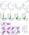
The endoU catalytic mutant H234A replicates less efficiently and elicits higher immune responses in the lungs of hamsters. Syrian hamsters were intranasally inoculated with 1 × 10³ PFU of WT virus (n = 8), H234A virus (n = 8), or with 100 µL of 1X PBS (n = 4). Half of the animals were killed on day 2 and the rest on day 5 postinfection, and their nasal turbinates, lungs, heart, and small intestine were harvested. (A) Viral titers were measured by the plaque assay on Caco-2/AT cells. (B) Expression of indicated genes in the lungs was measured by RT-qPCR. (C) Images of H&E-stained lung tissues collected at 5 dpi. Black arrows indicate foci of immune cell infiltrates. (D) The percent area of lung scans containing immune cell infiltrates was measured as detailed in the Materials and Methods section. Statistical significance was determined using the two-tailed parametric t test.
Fig. 5.
Fig. 5.
The endoU catalytic mutant H234A elicits increased accumulation of dsRNA in infected cells. (A) iAT2 cells infected with indicated viruses at an MOI of 0.3 were stained with anti-dsRNA antibody (green). Nuclei were counterstained with DAPI (blue). Representative images are shown. (B) Quantification of area per punctum. 5,640 puncta for WT virus and 12,809 puncta for H234A virus were quantified in two fields across 27-41 Z planes of iAT2 cells infected at an MOI of 0.3 for 48 h. (C) Gene expression in iAT2 cells infected at an MOI of 0.3 for 48 h in the presence or absence of 10 µM compounds was analyzed by RT-qPCR. (D) Virus titer in apical washes collected from iAT2 cells grown in the presence or absence of 10 µM compounds for indicated times was measured by the plaque assay on Caco-2/AT cells. Statistical significance was determined using the two-tailed parametric t test.

References

    1. Wolff G., et al. , A molecular pore spans the double membrane of the coronavirus replication organelle. Science 369, 1395–1398 (2020). - PMC - PubMed
    1. Ricciardi S., et al. , The role of NSP6 in the biogenesis of the SARS-CoV-2 replication organelle. Nature 606, 761–768 (2022). - PMC - PubMed
    1. Deng X., Baker S. C., An “Old” protein with a new story: Coronavirus endoribonuclease is important for evading host antiviral defenses. Virology 517, 157–163 (2018). - PMC - PubMed
    1. Zheng A., et al. , Insight into the evolution of nidovirus endoribonuclease based on the finding that nsp15 from porcine deltacoronavirus functions as a dimer. J. Biol. Chem. 293, 12054–12067 (2018). - PMC - PubMed
    1. Athmer J., et al. , In situ tagged nsp15 reveals interactions with coronavirus replication/transcription complex-associated proteins. MBio 8, e02320–02316 (2017). - PMC - PubMed

MeSH terms

LinkOut - more resources