. 2025 Sep;116(9):2580-2591.
doi: 10.1111/cas.70107. Epub 2025 Jun 12.

Implication of KLHL Gene Family Member KLHL5 in Colorectal Cancer Progression and Prognosis

Affiliations

Implication of KLHL Gene Family Member KLHL5 in Colorectal Cancer Progression and Prognosis

Konomu Uno et al. Cancer Sci. 2025 Sep.

Abstract

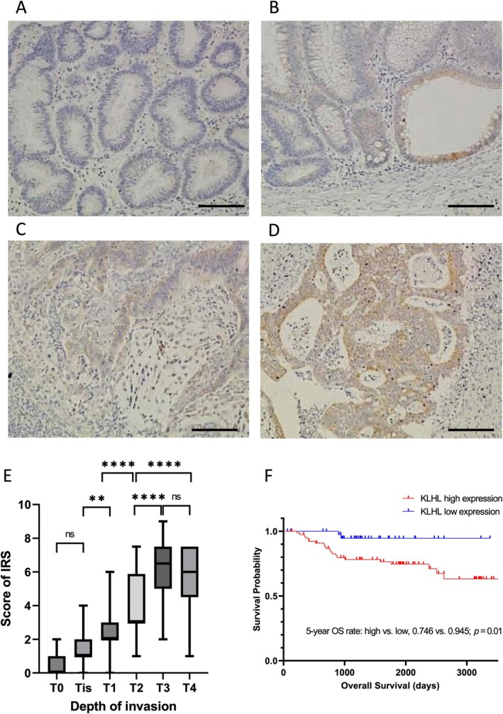
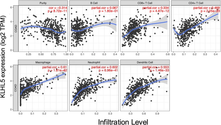
The KLHL gene family member KLHL5, which is a constituent factor of the RING E3 ubiquitin ligase complex, is expressed in various types of cancers and plays a role in cancer pathophysiology. In this study, we identified KLHL5 as a potential biomarker for predicting the prognosis of colorectal cancer (CRC) from 42 KLHL family genes using transcriptome profiles generated by RNA-seq analysis of The Cancer Genome Atlas colorectal adenocarcinoma (TCGA-COAD). We further investigated the implication of KLHL5 in CRC using pathological examination and bioinformatics analyses. Clinicopathological analyses revealed that KLHL5 was more highly expressed in CRC than in adjacent normal mucosa, and its expression level increased concomitantly with the CRC stage (p < 0.05). KLHL5 expression was associated with poor prognostic factors such as depth of invasion (p < 0.001), lymphovascular invasion (p = 0.029), and lymph node metastasis (p = 0.025). Notably, KLHL5 exhibited heterogeneous expression within the tumor, with pronounced expression observed at the invasive front of the tumor (p < 0.0001). Through bioinformatics analyses, we determined that elevated KLHL5 expression in CRC is significantly associated with poor prognosis. Furthermore, analysis from the Gene Expression Omnibus database indicated that KLHL5 expression was more pronounced in the common molecular subtype (CMS) 4 CRC, which is characterized as highly advanced, and the overall and recurrence-free survival rates were poor compared to other CMS groups. Our findings indicate that KLHL5 plays a pivotal role in the progression and development of CRC, and can be used as a potential biomarker and therapeutic target for CRC treatment.

Keywords: adenocarcinoma; biomarkers; colorectal neoplasms; pathology; transcriptome.


Conflict of interest statement

Shigeki Higashiyama is Associate Editor of Cancer Science. Other authors declare no conflicts of interest.

Figures

FIGURE 1
FIGURE 1
Identification of KLHL5 by bioinformatical approach using colorectal adenocarcinoma in the Cancer Genome Atlas. (A) Schema of narrowing down of KLHL family genes using TCGA‐COAD RNA‐seq data. Two KLHL family genes (KLHL5 and KLHL35) were extracted by transcriptome diversity approach using median and variation of gene expression. (B, C) Kaplan–Meier survival curves of disease‐free survival (B) and overall survival (C) for CRC patients with KLHL5 high and low expression were measured using PrognoScan. (D) Analysis of the correlation between KLHL5 expression and CMS in TCGA‐COAD RNA‐seq dataset. p‐values were calculated using the Kruskal–Wallis test with the Steel–Dwass post hoc test. *p < 0.05.
FIGURE 2
FIGURE 2
Immunohistochemical staining of KLHL5 in clinical samples and the quantification of KLHL5 expression in each stage of CRC. (A–D) Representative immunohistochemical images of KLHL5 staining in adenoma (A), T0 stage CRC (B), T1 stage CRC (C), and T2 stage CRC (D). Scale bar, 50 μm. (E) KLHL5 expression was quantified according to IRS in each stage of CRC (T1–T4). ns, not significant; **p < 0.01; ****p < 0.0001. (F) Kaplan–Meier survival curves of overall survival times, according to the expression level of KLHL5 in the advanced CRC (T2–T4).
FIGURE 3
FIGURE 3
Immunohistochemical staining of KLHL5 in the advanced CRC. (A) Immunohistochemical staining of KLHL5 in the advanced CRC. Scale bar, 1.0 mm. The magnified image of the central lesion of CRC (B) and the lower lesion of CRC (C). The boxes indicate the location of the magnified images. Scale bar, 100 μm. (D) Quantification of KLHL5 expression in the central lesion of the tumor and the lower lesion of the tumor. ****p < 0.0001.
FIGURE 4
FIGURE 4
Bioinformatic analyses of KLHL5 expression in CRC. (A, B) Expression levels of KLHL5 in normal colon and colorectal neoplastic tissues were analyzed using the Oncomine Cancer Profiling Database. (C) Immunoreactive score (IRS) of KLHL5 expression in matched tissue samples (adenoma and carcinoma) from patients with CRC. *p < 0.05, ****p < 0.0001.
FIGURE 5
FIGURE 5
Analysis of the correlation KLHL5 expression and CMS. (A‐I) The boxes represent the 25th, 50th (median), and 75th percentiles of KLHL5 mRNA expression. p‐values were calculated using the Kruskal–Wallis test with the Steel‐Dwass post hoc test. *p < 0.05, ***p < 0.001.
FIGURE 6
FIGURE 6
Immune cell infiltration with KLHL5 expression in the CRC patients. Estimation of Immune cell infiltration by TIMER algorithms with KLHL5 expression. Infiltrations of B cell, CD8+ T cell, CD4+ T cell, Macrophage, Neutrophil, and Dendritic cell were estimated. The purity‐corrected partial Spearman's rho value and statistical significance are shown in red.

References

    1. Sung H., Ferlay J., Siegel R. L., et al., “Global Cancer Statistics 2020: GLOBOCAN Estimates of Incidence and Mortality Worldwide for 36 Cancers in 185 Countries,” CA: A Cancer Journal for Clinicians 71, no. 3 (2021): 209–249, 10.3322/caac.21660. - DOI - PubMed
    1. Dhanoa B. S., Cogliati T., Satish A. G., Bruford E. A., and Friedman J. S., “Update on the Kelch‐Like (KLHL) Gene Family,” Human Genomics 7, no. 1 (2013): 13, 10.1186/1479-7364-7-13. - DOI - PMC - PubMed
    1. Adams J., Kelso R., and Cooley L., “The Kelch Repeat Superfamily of Proteins: Propellers of Cell Function,” Trends in Cell Biology 10, no. 1 (2000): 17–24, 10.1016/s0962-8924(99)01673-6. - DOI - PubMed
    1. Wang P., Song J., and Ye D., “CRL3s: The BTB‐CUL3‐RING E3 Ubiquitin Ligases,” Advances in Experimental Medicine and Biology 1217 (2020): 211–223, 10.1007/978-981-15-1025-0_13. - DOI - PubMed
    1. Deng L., Meng T., Chen L., Wei W., and Wang P., “The Role of Ubiquitination in Tumorigenesis and Targeted Drug Discovery,” Signal Transduction and Targeted Therapy 5, no. 1 (2020): 11, 10.1038/s41392-020-0107-0. - DOI - PMC - PubMed

Substances