

Expression landscape of epigenetic genes in human hepatocellular carcinoma
- PMID: 40504449
- PMCID: PMC12373563
- DOI: 10.1007/s13105-025-01095-6
Expression landscape of epigenetic genes in human hepatocellular carcinoma
Abstract
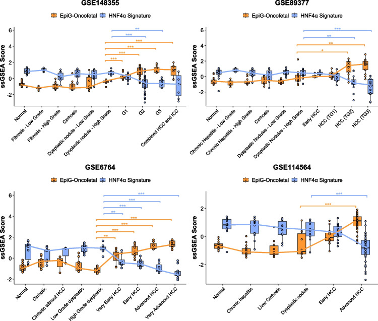
Hepatocellular carcinoma (HCC) is the most common primary liver tumor, often arising in the context of chronic liver disease. Despite recent advances in systemic therapies, including the use of immune checkpoint inhibitors (ICIs), clinical outcomes remain suboptimal, with many patients exhibiting primary or acquired resistance. Accumulating evidence indicates that the dysregulation of epigenetic mechanisms contributes to HCC development, and may also play a crucial role in shaping the tumor immune microenvironment, influencing responses to treatments. In this study, we analyzed the expression profiles of a comprehensive set of epigenetic regulators across publicly available transcriptomic datasets of HCC and non-tumoral liver tissues. Our findings reveal a consistent dysregulation of key epigenetic modifiers, particularly those involved in DNA methylation and histone modification. Furthermore, our analysis underscores the need for a deeper understanding of the epigenetic landscape of HCC, as specific epigenetic patterns are directly associated with disease development, the major mutational, immune, and transcriptional subclasses of HCC, and patient clinical outcomes. Our study provides a foundation for integrating epigenetic biomarkers into patient stratification and therapeutic decision-making. A more comprehensive analysis of epigenetic alterations could pave the way for novel predictive markers and combination strategies that could enhance the efficacy of ICIs in HCC.
Keywords: Epigenetics; Gene expression; Hepatocellular carcinoma; Immune landscape; Oncofetal reprogramming.
© 2025. The Author(s).
Conflict of interest statement
Declarations. Conflict of interest: The authors declare no competing interests. Clinical trial number: Not applicable.
Figures


References
-
- Ally A, Balasundaram M, Carlsen R, Chuah E, Clarke A, Dhalla N, Holt RA, Jones SJM, Lee D, Ma Y, Marra MA, Mayo M, Moore RA, Mungall AJ, Schein JE, Sipahimalani P, Tam A, Thiessen N, Cheung D, Wong T, Brooks D, Robertson AG, Bowlby R, Mungall K, Sadeghi S, Xi L, Covington K, Shinbrot E, Wheeler DA, Gibbs RA, Donehower LA, Wang L, Bowen J, Gastier-Foster JM, Gerken M, Helsel C, Leraas KM, Lichtenberg TM, Ramirez NC, Wise L, Zmuda E, Gabriel SB, Meyerson M, Cibulskis C, Murray BA, Shih J, Beroukhim R, Cherniack AD, Schumacher SE, Saksena G, Pedamallu CS, Chin L, Getz G, Noble M, Zhang H, Heiman D, Cho J, Gehlenborg N, Saksena G, Voet D, Lin P, Frazer S, Defreitas T, Meier S, Lawrence M, Kim J, Creighton CJ, Muzny D, Doddapaneni HV, Hu J, Wang M, Morton D, Korchina V, Han Y, Dinh H, Lewis L, Bellair M, Liu X, Santibanez J, Glenn R, Lee S, Hale W, Parker JS, Wilkerson MD, Hayes DN, Reynolds SM, Shmulevich I, Zhang W, Liu Y, Iype L, Makhlouf H, Torbenson MS, Kakar S, Yeh MM, Jain D, Kleiner DE, Jain D, Dhanasekaran R, El-Serag HB, Yim SY, Weinstein JN, Mishra L, Zhang J, Akbani R, Ling S, Ju Z, Su X, Hegde AM, Mills GB, Lu Y, Chen J, Lee JS, Sohn BH, Shim JJ, Tong P, Aburatani H, Yamamoto S, Tatsuno K, Li W, Xia Z, Stransky N, Seiser E, Innocenti F, Gao J, Kundra R, Zhang H, Heins Z, Ochoa A, Sander C, Ladanyi M, Shen R, Arora A, Sanchez-Vega F, Schultz N, Kasaian K, Radenbaugh A, Bissig KD, Moore DD, Totoki Y, Nakamura H, Shibata T, Yau C, Graim K, Stuart J, Haussler D, Slagle BL, Ojesina AI, Katsonis P, Koire A, Lichtarge O, Hsu TK, Ferguson ML, Demchok JA, Felau I, Sheth M, Tarnuzzer R, Wang Z, Yang L, Zenklusen JC, Zhang J, Hutter CM, Sofia HJ, Verhaak RGW, Zheng S, Lang F, Chudamani S, Liu J, Lolla L, Wu Y, Naresh R, Pihl T, Sun C, Wan Y, Benz C, Perou AH, Thorne LB, Boice L, Huang M, Rathmell WK, Noushmehr H, Saggioro FP, da Tirapelli DPC, Junior CGC, Mente ED, de Silva OC, Trevisan FA, Kang KJ, Ahn KS, Giama NH, Moser CD, Giordano TJ, Vinco M, Welling TH, Crain D, Curley E, Gardner J, Mallery D, Morris S, Paulauskis J, Penny R, Shelton C, Shelton T, Kelley R, Park JW, Chandan VS, Roberts LR, Bathe OF, Hagedorn CH, Auman JT, O’Brien DR, Kocher JPA, Jones CD, Mieczkowski PA, Perou CM, Skelly T, Tan D, Veluvolu U, Balu S, Bodenheimer T, Hoyle AP, Jefferys SR, Meng S, Mose LE, Shi Y, Simons JV, Soloway MG, Roach J, Hoadley KA, Baylin SB, Shen H, Hinoue T, Bootwalla MS, Van Den Berg DJ, Weisenberger DJ, Lai PH, Holbrook A, Berrios M, Laird PW (2017) Comprehensive and Integrative Genomic Characterization of Hepatocellular Carcinoma. Cell 169:1327-1341.e23. 10.1016/j.cell.2017.05.046 - PMC - PubMed
-
- Avila MA, Berasain C, Torres L, Martín-Duce A, Corrales FJ, Yang H, Prieto J, Lu SC, Caballería J, Rodés J, Mato JM (2000) Reduced mRNA abundance of the main enzymes involved in methionine metabolism in human liver cirrhosis and hepatocellular carcinoma. J Hepatol 33:907–914. 10.1016/S0168-8278(00)80122-1 - PubMed
-
- Ayers M, Lunceford J, Nebozhyn M, Murphy E, Loboda A, Kaufman DR, Albright A, Cheng JD, Kang SP, Shankaran V, Piha-Paul SA, Yearley J, Seiwert TY, Ribas A, McClanahan TK (2017) IFN-γ-related mRNA profile predicts clinical response to PD-1 blockade. J Clin Invest 127:2930–2940. 10.1172/JCI91190 - PMC - PubMed
MeSH terms
Substances
Grants and funding
- CD22/00109/Sara Borrell Contract From Spanish Ministry of Health
- TRANSCAN2022-784-024/European Union Horizon 2020 research and innovation programme. ERANET-TRANSCAN.
- FPU23/00176/MINISTERIO DE CIENCIA, INNOVACIÓN Y UNIVERSIDADES, Programa de Formación del Profesorado Universitario (FPU)
- INVES223049AREC/AECC investigador fellowship
- PID2022-136616OB-I00/AEI/10.13039/501100011033/Ministerio de Ciencia Innovación y Universidades MICINN-Agencia Estatal de Investigación integrado en el Plan Estatal de Investigación Científica y Técnica y Innovación, cofinanciado con Fondos FEDER
- PID2020-117116RB-I00/Ministerio de Ciencia Innovación y Universidades MICINN-Agencia Estatal de Investigación integrado en el Plan Estatal de Investigación Científica y Técnica y Innovación, cofinanciado con Fondos FEDER
- LABAE20011GARC/Scientific Foundation of the Spanish Association Against Cancer (AECC)
- RYC2018-024475-1/Ramón y Cajal Program contract
LinkOut - more resources
Full Text Sources
Medical
