. 2025 Jun 12;23(6):e3002738.
doi: 10.1371/journal.pbio.3002738. eCollection 2025 Jun.

Global siRNA screen identifies human host factors critical for SARS-CoV-2 replication and late stages of infection

Affiliations

Global siRNA screen identifies human host factors critical for SARS-CoV-2 replication and late stages of infection

Xin Yin et al. PLoS Biol. .

Abstract

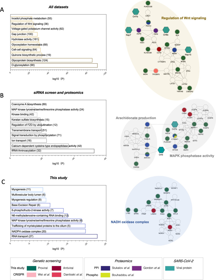
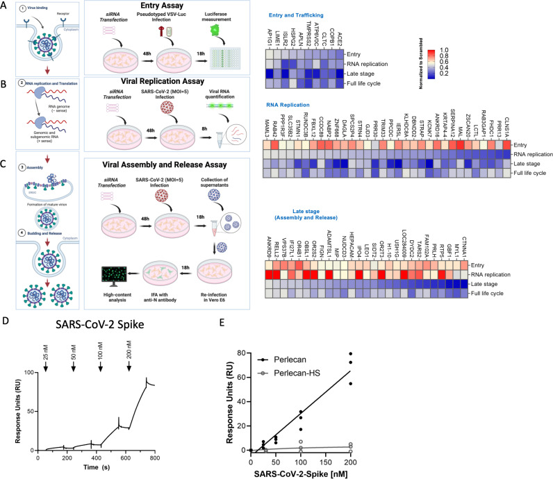
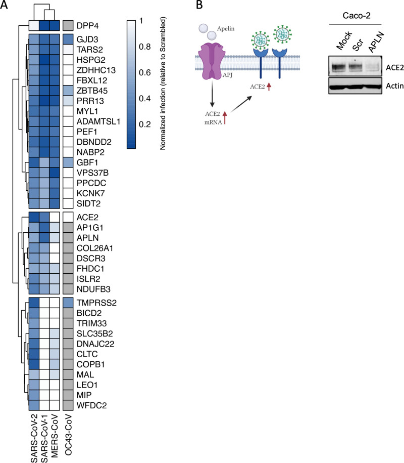
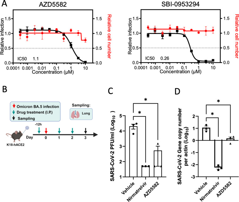
Defining the subset of cellular factors governing SARS-CoV-2 replication can provide critical insights into viral pathogenesis and identify targets for host-directed antiviral therapies. While a number of genetic screens have previously reported SARS-CoV-2 host dependency factors, most of these approaches relied on utilizing pooled genome-scale CRISPR libraries, which are biased toward the discovery of host proteins impacting early stages of viral replication. To identify host factors involved throughout the SARS-CoV-2 infectious cycle, we conducted an arrayed genome-scale siRNA screen. Resulting data were integrated with published functional screens and proteomics data to reveal (i) common pathways that were identified in all OMICs datasets-including regulation of Wnt signaling and gap junctions, (ii) pathways uniquely identified in this screen-including NADH oxidation, or (iii) pathways supported by this screen and proteomics data but not published functional screens-including arachionate production and MAPK signaling. The identified proviral host factors were mapped into the SARS-CoV-2 infectious cycle, including 32 proteins that were determined to impact viral replication and 27 impacting late stages of infection, respectively. Additionally, a subset of proteins was tested across other coronaviruses revealing a subset of proviral factors that were conserved across pandemic SARS-CoV-2, epidemic SARS-CoV-1 and MERS-CoV, and the seasonal coronavirus OC43-CoV. Further studies illuminated a role for the heparan sulfate proteoglycan perlecan in SARS-CoV-2 viral entry and found that inhibition of the non-canonical NF-kB pathway through targeting of BIRC2 restricts SARS-CoV-2 replication both in vitro and in vivo. These studies provide critical insight into the landscape of virus-host interactions driving SARS-CoV-2 replication as well as valuable targets for host-directed antivirals.


Conflict of interest statement

I have read the journal's policy and the authors of this manuscript have the following competing interests: J.F.H. has received research support, paid to Northwestern University, from Gilead Sciences, and is a paid consultant for Merck. The A.G.-S. laboratory has received research support from GSK, Pfizer, Senhwa Biosciences, Kenall Manufacturing, Blade Therapeutics, Avimex, Johnson & Johnson, Dynavax, 7Hills Pharma, Pharmamar, ImmunityBio, Accurius, Nanocomposix, Hexamer, N-fold LLC, Model Medicines, Atea Pharma, Applied Biological Laboratories and Merck, outside of the reported work. A.G.-S. has consulting agreements for the following companies involving cash and/or stock: Castlevax, Amovir, Vivaldi Biosciences, Contrafect, 7Hills Pharma, Avimex, Pagoda, Accurius, Esperovax, Applied Biological Laboratories, Pharmamar, CureLab Oncology, CureLab Veterinary, Synairgen, Paratus, Pfizer and Prosetta, outside of the reported work. A.G.-S. has been an invited speaker in meeting events organized by Seqirus, Janssen, Abbott, Astrazeneca, and Novavax. A.G.-S. is inventor on patents and patent applications on the use of antivirals and vaccines for the treatment and prevention of virus infections and cancer, owned by the Icahn School of Medicine at Mount Sinai, New York, outside of the reported work. T.I. is cofounder of Data4Cure, is on the Scientific Advisory Board, and has an equity interest. T.I. is on the Scientific Advisory Board of Ideaya BioSciences and has an equity interest. The terms of these arrangements have been reviewed and approved by the University of California San Diego, in accordance with its conflict of interest policies. All other authors declare no competing interests.

Figures

Fig 1
Fig 1. Genome-wide siRNA screen identifies host factors involved in SARS-CoV-2 replication.
(A) Schematic representation of the genome-wide screen to identify human host factors that affect SARS-CoV-2 replication. (B) Ranked SARS-CoV-2 infectivity Z-scores from the genome-wide siRNA screen. Dashed lines illustrate cut-offs for hit calling strategy: Z-score ≤ −2 indicates proviral factors (green), Z-score ≥ 1.5 indicates antiviral factors (red). Controls are shown (e.g., siACE2, positive). (C) Functional enrichment analysis of identified proviral (left-green) and antiviral (right-red) host factors. (D) Deconvolution plot showing proviral host factors validated with one siRNA (gray), two siRNAs (dark blue), three siRNAs (light blue), and four siRNAs (pink). (E) Calu-3 cells treated with indicated gRNAs were infected with SARS-CoV-2 (MOI = 0.75) for 48 h prior to immunostaining for viral N protein. Shown is quantification of the normalized infection (% of SARS-CoV-2 N+ cells) relative to parental cells. Data show mean ± SD from one representative experiment in quadruplicate (n = 4) of two independent experiments. Significance was calculated using one-way ANOVA with Dunnett’s post-hoc test. The data underlying this figure can be found in S1 Data.
Fig 2
Fig 2. Network integration reveals relevant networks implicated in SARS-CoV-2 replication.
The network containing the identified proviral (green) and antiviral (red) human host factors was integrated with host factors reported to be relevant for SARS-CoV-2 infection. These include genetic CRISPR screen hits (Wei and colleagues, 2020 [15], light pink; Daniloski and colleagues, 2020 [16], dark pink), protein–protein interaction hits (Stukalov and colleagues, 2020 [31], blue; Gordon and colleagues, 2020 [32], purple), as well as hits from a phosphoproteomics study (Bouhaddou and colleagues, 2020 [33], yellow). The network was subjected to supervised community detection [67,73], and then clustered based on identification by all datasets (A), this screen and proteomics data but not CRISPR screens (B), or exclusively by this screen (C). The top 10 GO categories with the lowest p-values are shown. Continuous black edges indicate interactions from STRING database, discontinued edges indicate virus-host interactions. Turquoise nodes indicate SARS-CoV-2 proteins. White denotes proteins in network (based on STRING) but not identified in any of the OMICs studies. * indicates highlighted clusters.
Fig 3
Fig 3. Mapping of host factors into the SARS-CoV-2 replication cycle reveals a direct interaction between entry factor perlecan and SARS-CoV-2 S protein.
(A) Caco-2 cells were subjected to siRNA-mediated knockdown of indicated host factors and then infected with SARS-CoV-2 pseudotyped VSV luciferase virus (VSV-S-luc) for 18 h prior to measurement of luciferase signal. (B) In parallel, cells were subjected to synchronized infection with SARS-CoV-2 (MOI = 5) for 8 h prior to measurement of viral RNA, or (C) supernatants collected at 18 h post-infection were used to infect naïve Vero E6 cells. The % of infected cells was then determined at 18 h post-infection using immunostaining for viral N protein (3−4). In parallel to these experiments, the impact of depleting these factors on SARS-CoV-2 replication was evaluated at 24 h post-infection in Caco-2 cells (full replication cycle, Fig 3A–3C). Results are summarized in the heat map and show the mean (n = 2) of relative activities compared to cells treated with non-targeting scramble siRNA. (D, E) Surface plasmon resonance (SPR) was used to evaluate the binding of S protein to perlecan or perlecan without HS binding to immunopurified perlecan isolated from human coronary artery endothelial cells. Control flow channels contained immobilized BSA. S protein at indicated concentrations was run across the flow channels for 120 s and dissociation was measured in the following 600 s. The RU values throughout the experiment for BSA were subtracted from the RU values for perlecan to determine the level of specific binding. This experiment was repeated with perlecan treated with heparinase III. The data underlying this figure can be found in S1 Data.
Fig 4
Fig 4. Comparative screening reveals proviral factors that are conserved across several Coronaviruses.
(A) Heat map showing normalized infection of SARS-CoV-1, CoV-2, MERS-CoV, and OC43-CoV upon knockdown of indicated human host factors. Caco-2 cells depleted for indicated factors were infected with SARS-CoV-2 (MOI = 0.625) for 48 h prior to immunostaining for viral N protein. Shown is quantification of the normalized infection (% of SARS-CoV-2 N+ cells) relative to control cells (scrambled siRNA). A549-DPP4, A549-ACE2, or A549 were depleted for indicated factors and then infected with MERS-CoV, SARS-CoV-1, or OC43-CoV respectively (MOI 0.1). At 48 h post-infection, supernatants were collected and used to calculate the TCID50. Data shows TCID50/ml relative to control cells (scrambled siRNA). Data show mean ± SD from one representative experiment in duplicate (n = 2) of two independent experiments. Cells in gray mean the factor was not tested. (B) Cell lysates from Caco-2 cells mock-treated or treated with scrambled or APLN siRNAs for 48 h were then subjected to SDS-PAGE and immunoblotted using antibodies specific for ACE2 and Actin (loading control). Blot is representative of two independent experiments. The data underlying this figure can be found in S1 Data. The original uncropped blots can be found in S1 Raw Images.
Fig 5
Fig 5. Pharmacological inhibition of BIRC2 reduces SARS-CoV-2 replication in vitro and in vivo.
(A) Dose–response analysis of SBI-0953294 and AZD5582 showing infectivity (black), cell number (red), and cellular IC50 values. (B) Layout of mice experiments. Effect of AZD5582 on SARS-CoV-2 Omicron replication in the lungs of infected mice as measured by plaque assay (C) and qRT-PCR (D). Tissue sampling was done at 72 hpi. One-way ANOVA when compared with the vehicle control group, *p < 0.05. And the detection limit = 50 PFU/ml in a 12-well plate. The data underlying this figure can be found in S1 Data.

Update of

References

    1. Zhu Z, Lian X, Su X, Wu W, Marraro GA, Zeng Y. From SARS and MERS to COVID-19: a brief summary and comparison of severe acute respiratory infections caused by three highly pathogenic human coronaviruses. Respir Res. 2020;21(1):224. doi: 10.1186/s12931-020-01479-w - DOI - PMC - PubMed
    1. Abdelrahman Z, Li M, Wang X. Comparative review of SARS-CoV-2, SARS-CoV, MERS-CoV, and influenza A respiratory viruses. Front Immunol. 2020;11:552909. doi: 10.3389/fimmu.2020.552909 - DOI - PMC - PubMed
    1. Carabelli AM, Peacock TP, Thorne LG, Harvey WT, Hughes J, COVID-19 Genomics UK Consortium, et al. SARS-CoV-2 variant biology: immune escape, transmission and fitness. Nat Rev Microbiol. 2023;21(3):162–77. doi: 10.1038/s41579-022-00841-7 - DOI - PMC - PubMed
    1. The evolution of SARS-CoV-2 | Nature Reviews Microbiology https://www.nature.com/articles/s41579-023-00878-2. - PubMed
    1. Kaufmann SHE, Dorhoi A, Hotchkiss RS, Bartenschlager R. Host-directed therapies for bacterial and viral infections. Nat Rev Drug Discov. 2018;17(1):35–56. doi: 10.1038/nrd.2017.162 - DOI - PMC - PubMed

Substances