. 2025 Jun 12;388(6752):1218-1224.
doi: 10.1126/science.ado6670. Epub 2025 Jun 12.

Pan-viral ORFs discovery using massively parallel ribosome profiling

Affiliations

Pan-viral ORFs discovery using massively parallel ribosome profiling

Shira Weingarten-Gabbay et al. Science. .

Abstract

Defining viral proteomes is crucial to understanding viral life cycles and immune recognition but the landscape of translated regions remains unknown for most viruses. We have developed massively parallel ribosome profiling (MPRP) to determine open reading frames (ORFs) across tens of thousands of designed oligonucleotides. MPRP identified 4208 unannotated ORFs in 679 human-associated viral genomes. We found viral peptides originating from detected noncanonical ORFs presented on class-I human leukocyte antigen in infected cells and hundreds of upstream ORFs that likely modulate translation initiation of viral proteins. The discovery of viral ORFs across a wide range of viral families-including highly pathogenic viruses-expands the repertoire of vaccine targets and reveals potential cis-regulatory sequences.


Conflict of interest statement

Competing interests: S.W.-G., M.R.B, A.C.S., and P.C.S. are named co-inventors on International Patent Application No. PCT/US2024/048478 Claiming priority to U.S. Provisional Application No. 63/540,279 related to this work filed by The Broad Institute that covers massively parallel methods and techniques for evaluating open reading frames in genomes, particularly viral genomes. These methods and data unveil new proteins, immune targets, and cis-regulatory elements that can serve in the design of vaccines and commercial overexpression platforms.. M.R.B is an employee of BioNTech SE. S.K is an employee of Genentech. S.A.C. is a member of the scientific advisory boards of Kymera, PTM BioLabs, Seer and PrognomIQ. JGA is a paid consultant for Enara Bio and Moderna. P.C.S. is a co-founder of and consultant to Sherlock Biosciences and Delve Biosciences, is on the Board of Directors and a shareholder of Danaher Corporation and Polaris Genomics and holds equity in all the companies. The remaining authors declare no competing interests. All other authors declare no competing interests.

Figures

Figure 1.
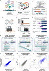
Figure 1.. Design of oligonucleotide synthetic library and MPRP measurements
(A) Illustration of the Massively Parallel Ribosome Profiling experiment (MPRP). (i) Synthetic library amplification using constant primers. (ii) Cloning library into overexpression vector. (iii) Transient transfection of plasmid pool into HEK293T or A549 cells for 24 h. (iv) Treating cells with either LTM or CHX and performing ribosome profiling protocol. (v) Mapping deep sequencing reads to the synthetic library. (vi) Inferring translated ORFs using PRICE (19). (B) Design of the tested synthetic oligonucleotides: (i) ORFs identified by ribosome profiling in infected cells with either intact start codon or a GCC mutation. (ii) Tiling oligos encompassing complete viral transcripts. (iii) Oligos spanning the 5’UTR and the first 140 nt of annotated viral CDSs. For the region containing the CDS, two oligos were designed: the wild-type sequence and a start codon mutated oligo. (C) Comparing the number of ribosome footprints mapped to 15,000 oligos in two biological replicates in HEK293T cells. R=0.92, Pearson correlation. (D) Comparing the number of ribosome footprints mapped to 15,000 oligos in HEK293T and A549 cells. R=0.89, Pearson correlation. Showing in color oligos with shared sequences with the adenoviral E1A/B genes and the SV40 large T-antigen endogenously expressed by HEK293T cells. (E) Comparing the number of ribosome footprints mapped to 1,163 identical oligos in two synthetic libraries in HEK293T cells. R=0.79, Pearson correlation.
Figure 2.
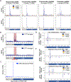
Figure 2.. Annotated CDSs measurements in MPRP and infected cells
(A) Metagene analysis for four viral families with CHX and LTM inhibitors showing the average ribosome footprints in each position. See Figures S6 and S7 for 21 viral families. (B) Comparing the average number of ribosome footprints between oligos containing the wt start codon (upper graph) to those in which the annotated start codon was mutated to GCC (lower graph). Shown are the average ribosome footprints in each position across 3,777 oligos. (C) ORF discovery using PRICE. Showing the number of ORFs detected in each position (ORF start position). (D) Mirror plot showing the number of ribosome footprints mapped to the first 200nt of the Nucleocapsid transcript in VSV-infected cells and MPRP. (E) Similar to (D) for IAV NEP and PB1 transcripts.
Figure 3.
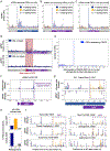
Figure 3.. Non-canonical ORFs measurements in MPRP and infected cells
(A-C) Metagene analysis of oligos containing the sequence of ORFs that were identified by ribosome profiling of HCMV-infected cells (20). Showing the average of ribosome footprints in each position. (D) Comparing the average number of ribosome footprints between oligos containing the wt start codon (upper graph) to those in which the reported start codon was mutated to GCC (lower graph). Shown are the average ribosome footprints across 284 oligos, containing Ribo-seq ORFs in the length of 7–45 aa. (E) ORF discovery using PRICE. Showing the number of ORFs detected in each position (ORF start position). (F) Mirror plot showing the number of ribosome footprints in HCMV-infected cells and MPRP. Purple box highlights uORF2, which encodes a ribosome arrest peptide (21) (G) Mirror plot showing the number of ribosome footprints in IAV-infected cells and MPRP. The purple box highlights an internal overlapping iORF in the +1 reading frame. (H) Percentages of ribosome footprints mapped to 0, +1, and −1 reading frames in the region encoding the internal ORF (upper panel) and outside this region (lower panel). (I) Ribosome footprints on the M1 coding sequences of six additional IAV strains from the MPRP experiment.
Figure 4.
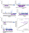
Figure 4.. HLA-I peptides derived from non-canonical ORFs in HCMV and VACV
(A) HLA-I peptides detected in four non-canonical ORFs of HCMV identified by MPRP. (B) HLA-I peptides originating from two non-canonical ORFs in VACV: A uORF in the non-coding region upstream of the I7L coding sequence, and an iORF overlapping the coding region of L3L. (C) Comparing HLA-I presentation from annotated and non-canonical ORFs in HCMV. For each ORF, we present the number of total HLA-I peptides detected in HCMV immunopeptidome (not unique) normed by the ORF length. p<10−3, Wilcoxon rank-sum test.
Figure 5.
Figure 5.. Ribosome densities on uORFs and CDSs in response to eIF2alpha phosphorylation
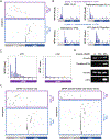
(A) Heatmap showing ribosome footprint densities across 2,418 viral oligos. Each line represents a single viral gene and each column represents the position relative to the annotated start codon. Genes in the upper cluster (purple) had more footprints in the 5’UTR region than the CDS region, and genes in the lower cluster (blue) had more footprints in the CDS than the 5’UTR. (B) Example of two individual genes from each cluster and the distribution of ribosome footprints observed in each position. (C) Metagene analysis showing the average ribosome footprints in each position along uORFs detected by PRICE, relative to the uORF start position. Shown for CHX (left) and LTM (right) inhibitors. (D) Western blot analysis of lysates from HEK293T cells treated with 40uM sodium-arsenite for 30 and 60 min. Phosphorylated eIF2alpha was detected with a monoclonal phospho S51 antibody (upper panel). ATF4 protein was detected using a polyclonal antibody (lower panel). In both membranes, Vinculin was used as a loading control. (E) Repeating the MPRP experiment in HEK293T cells that were treated with 40uM sodium arsenite and in non-treated cells. Shown are heatmaps of ribosome densities across viral oligos and clusters similarly to the analysis in (A).

Update of

References

    1. Lulla V, Dinan AM, Hosmillo M, Chaudhry Y, Sherry L, Irigoyen N, Nayak KM, Stonehouse NJ, Zilbauer M, Goodfellow I, Firth AE, An upstream protein-coding region in enteroviruses modulates virus infection in gut epithelial cells. Nat Microbiol 4, 280–292 (2019). - PMC - PubMed
    1. Ogden PJ, Kelsic ED, Sinai S, Church GM, Comprehensive AAV capsid fitness landscape reveals a viral gene and enables machine-guided design. Science 366, 1139–1143 (2019). - PMC - PubMed
    1. Lulla V, Firth AE, A hidden gene in astroviruses encodes a viroporin. Nat. Commun. 11, 4070 (2020). - PMC - PubMed
    1. Bansal A, Carlson J, Yan J, Akinsiku OT, Schaefer M, Sabbaj S, Bet A, Levy DN, Heath S, Tang J, Kaslow RA, Walker BD, Ndung’u T, Goulder PJ, Heckerman D, Hunter E, Goepfert PA, CD8 T cell response and evolutionary pressure to HIV-1 cryptic epitopes derived from antisense transcription. J. Exp. Med. 207, 51–59 (2010). - PMC - PubMed
    1. Ingolia NT, Brar GA, Stern-Ginossar N, Harris MS, Talhouarne GJS, Jackson SE, Wills MR, Weissman JS, Ribosome profiling reveals pervasive translation outside of annotated protein-coding genes. Cell Rep. 8, 1365–1379 (2014). - PMC - PubMed

LinkOut - more resources