. 2025 Sep:67:101194.
doi: 10.1016/j.neo.2025.101194. Epub 2025 Jun 11.

ACSL4 knockdown inhibits colorectal cancer progression through stimulating anti-tumor immunity

Affiliations

ACSL4 knockdown inhibits colorectal cancer progression through stimulating anti-tumor immunity

Yiming Liu et al. Neoplasia. 2025 Sep.

Abstract

Background: Long-chain acyl-CoA synthetase 4 (ACSL4), a crucial modulator of ferroptosis, is associated with tumor progression, though its impact on colorectal cancer (CRC) immune dynamics is not fully understood.

Methods: ACSL4 expression was analyzed in CRC tissues and correlated with patient prognosis. Effects of ACSL4 were evaluated in CRC cells in vitro and in subcutaneous and orthotopic CRC models. Flow cytometry and immunofluorescence were used to evaluate immune cell infiltration. RNA sequencing and RT-PCR were employed to identify ACSL4-regulated signaling pathways. The effect of ACSL4 silencing on PD-L1 blockade efficacy was also examined.

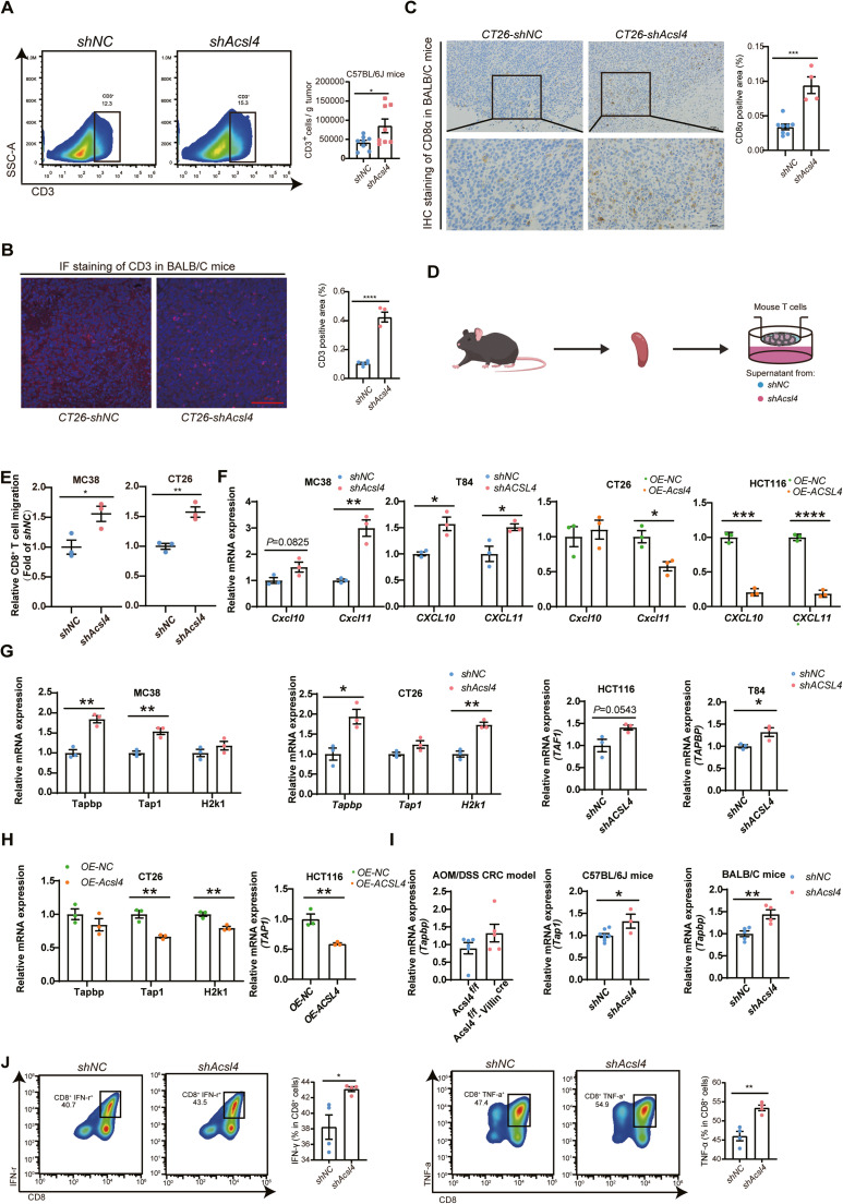
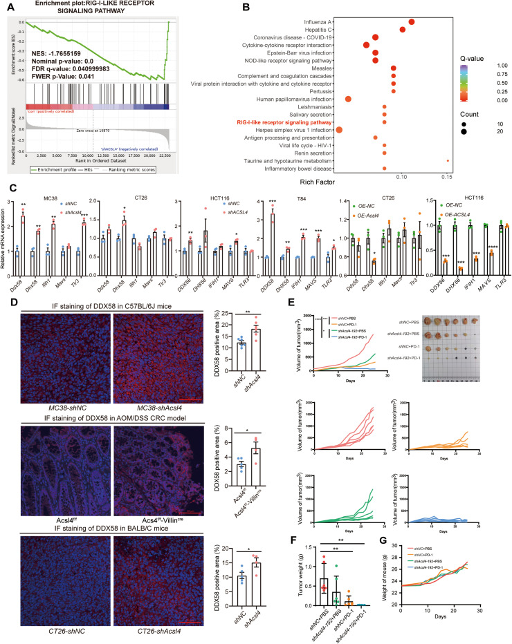
Results: ACSL4 levels were markedly increased in CRC and linked to unfavorable patient outcomes. While ACSL4 knockdown had no direct effect on CRC cell proliferation, it significantly suppressed tumor growth in immunocompetent mice. ACSL4 depletion enhanced CD3⁺ and CD8⁺ T cell infiltration and upregulated chemokines (CXCL10, CXCL11) and antigen presentation genes (H2k1, TAP1, TAPBP). Transcriptomic analysis highlighted activation of the RIG-I-MAVS-driven type I interferon pathway. Co-culture assays demonstrated that ACSL4 knockdown promoted CD8⁺ T cell activation, and ACSL4-deficient tumors exhibited enhanced responsiveness to PD-L1 blockade.

Conclusions: ACSL4 suppresses anti-tumor immunity in CRC by modulating the RIG-I-MAVS-IFN pathway, highlighting ACSL4 as a promising target for CRC immunotherapy.

Keywords: ACSL4; Colorectal cancer; RIG-I-MAVS; Tumor immunity; Type I Interferon.


Conflict of interest statement

Declaration of competing interest The authors declare that they have no known competing financial interests or personal relationships that could have appeared to influence the work reported in this paper.

Figures

Image, graphical abstract
Graphical abstract
Fig 1
Fig. 1
ACSL4 Overexpression in CRC Correlates with Poor Prognosis. (A) Expression levels of ACSL4 in CRC patients from the GSE156451 and GSE146889 datasets in the GEO database (GSE156451: Normal = 85, Tumor = 91; GSE146889: Normal = 72, Tumor = 72); (B) ACSL4 expression in CRC from the GEPIA database (Tumor = 275, Normal = 41); (C) ACSL4 expression levels in 21 sessile serrated adenomas/polyps (SSA/P), 10 hyperplastic polyps (HP), 10 adenomatous polyps (AP), 21 unaffected colon samples (UL, UR), 20 control colon samples (CL, CR), and 4 Colon Cancer (CA) samples from the GEO database. (D) RT-PCR analysis of Acsl4 expression in DSS-induced APCmin/+ mice from tumor nests (T) and adjacent tissues (P). (E) Immunohistochemical staining of Acsl4 in colorectal tissues from DSS-induced APCmin/+ mice (40×Scale bar: 200 μm, 100×Scale bar: 100 μm). (F) Statistical analysis of Acsl4 expression in colorectal tissues from DSS-induced APCmin/+ mice. (G) The relationship between ACSL4 expression and patient survival was analyzed using the PROGgeneV2 database. Survival analysis of CRC patients with ACSL4 expressions from the GSE29621, GSE14333, and GSE17536 datasets (GSE29621: High = 33, Low = 32; GSE14333: High = 94, Low = 93; GSE17536: High = 87, Low = 87). Data are expressed as the mean ± SD (n = 6 – 9) in B, D, and F. *P < 0.05, **P < 0.01, ****P < 0.0001.
Fig 2
Fig. 2
Knockdown of ACSL4 Inhibits Tumor Progression by Enhancing Anti-Tumor Immunity in Immunocompetent Mice. (A) SRB assay measuring in vitro proliferation of stable ACSL4 knockdown cell lines (MC38, CT26, HCT116 and T84) and stable Acsl4 overexpression cell line (CT26). (B) Growth curves of tumors implanted subcutaneously with Acsl4 knockdown MC38 cells; images of subcutaneous tumors at the experimental endpoint and corresponding tumor weight statistics; (C) Growth curves of tumors implanted subcutaneously with Acsl4 knockdown CT26 cells; images of subcutaneous tumors at the experimental endpoint and corresponding tumor weight statistics. (D) Immunohistochemical analysis and quantification of Ki-67 staining in subcutaneous MC38 tumors with Acsl4 knockdown (200×Scale bar: 50 μm); immunohistochemical analysis and quantification of PCNA staining in subcutaneous MC38 tumors with Acsl4 knockdown (200×Scale bar: 50 μm); immunohistochemical analysis and quantification of Ki-67 staining in subcutaneous CT26 tumors with Acsl4 knockdown (200×Scale bar: 50 μm). (E) Schematic of the CRC model in AOM/DSS-induced Acsl4f/f-Villincre mice; representative images of dissected colorectal tissues and H&E staining, with tumor statistics at the experimental endpoint; (F) Tumor growth curves of BALB/c-nude mice; Representative images of subcutaneous tumors and tumor weight statistics at the experimental endpoint for BALB/c-nude mice; (G) Tumor growth curves of C57BL/6 J mice; Representative images of subcutaneous tumors and tumor weight statistics at the experimental endpoint for C57BL/6 J mice; (H) Comparison of tumor inhibition rates between nude mice and C57BL/6 J mice. Data are expressed as the mean ± SD (n = 3–10). *P < 0.05, **P < 0.01, ***P < 0.001, ****P < 0.0001.
Fig 3
Fig. 3
Impact of ACSL4 Modulation on Colorectal Cancer Progression and Tumor Growth. (A) Flow cytometry analysis and statistical data showing the infiltration of CD3+T cells in subcutaneous tumors of Acsl4-knockdown MC38 cells. (B) Immunofluorescence staining images and statistical data of CD3 in subcutaneous tumor tissues from CT26 Acsl4 knockdown mice (200×Scale bar:50 μm); (C) Immunohistochemical staining and statistical data of CD8α expression in subcutaneous tumors from CT26 Acsl4 knockdown mice (200×Scale bar:50 μm, 400×Scale bar: 20 μm). (D) Schematic diagram of the in vitro T-cell chemotaxis assay; (E) Statistical data of the chemotaxis assay using conditioned medium from Acsl4-knockdown MC38 and CT26 cells; (F) RT-PCR analysis of CXCL10 and CXCL11 expression in ACSL4-knockdown MC38, T84, and ACSL4-overexpressing CT26 and HCT116 cells. (G) RT-PCR analysis of Tapbp, Tap1, and H2k1 expression in Acsl4-knockdown MC38 and CT26 cells; TAF1 expression in ACSL4-knockdown HCT116 cells; TAPBP expression in ACSL4-knockdown T84 cells; (H) Tapbp, Tap1, H2k1 expression in ACSL4-overexpressing CT26 and HCT116 cells; (I) RT-PCR analysis of Tapbp expression in AOM/DSS-induced Acsl4f/f-Villincre mice colorectal tissues; Tap1 expression in Acsl4-knockdown MC38 subcutaneous tumors; Tapbp expression in Acsl4-knockdown CT26 subcutaneous tumors. (J) Flow cytometry analysis and statistical data of T-cell effector cytokine secretion. Data are expressed as the mean ± SD (n = 3 – 8). *P < 0.05, **P < 0.01, ***P < 0.001, ****P < 0.0001.
Fig 4
Fig. 4
ACSL4 knockdown enhances type I interferon signaling and anti-tumor immunity in colorectal cancer cells through upregulation of the RIG-I-MAVS pathway. (A) Volcano plot showing differentially expressed genes in RNA-seq data from stable Acsl4-knockdown MC38 cells; (B) GO enrichment scatter plot of genes with differential expression in RNA-seq data from stable Acsl4-knockdown MC38 cells; (C) GeneRatio enrichment scatter plot of differentially expressed genes in RNA-seq data from stable ACSL4-knockdown T84 cells; (D) RT-PCR analysis of IRF7, IRF9, STAT1, STAT2 expression in stable ACSL4-knockdown MC38, CT26, HCT116, and T84 cells, as well as in ACSL4-overexpressing CT26 and HCT116 cells. (E) WB analysis of the impact of ACSL4 knockdown on type I IFN signaling pathways in MC38 and T84 cells. (F) RT-PCR analysis of IFN-β1 expression in CRC cells with stable ACSL4 knockdown or overexpression. (G) RT-PCR analysis of ISG expression in CRC cells with stable ACSL4 knockdown or overexpression. Data are expressed as the mean ± SD (n = 3). *P < 0.05, **P < 0.01, ***P < 0.001.
Fig 5
Fig. 5
ACSL4 knockdown activates the RIG-I-MAVS pathway, enhancing anti-tumor immunity in CRC. (A) KEGG pathway enrichment analysis of RNA-seq data from stable Acsl4-knockdown MC38 cells. (B) GSEA enrichment scatters plot of differentially expressed genes in RNA-seq data from stable Acsl4-knockdown MC38 cells. (C) RT-PCR analysis to evaluate the effect of ACSL4 expression on the RIG-I-like receptor and MAVS pathways in CRC cells; (D) Immunofluorescence staining and statistical analysis of DDX58 in subcutaneous tumor tissues from MC38 Acsl4-knockdown mice, CT26 Acsl4-knockdown mice, and AOM/DSS-induced CRC tissues in Acsl4f/f-Villincre mice (200×, Scale bar: 50 μm); (E) Growth curves of CT26 tumors in BALB/c nude mice with Acsl4 knockdown; (F) Tumor weight (g) of BALB/c-nude mice, along with the weight of the mice (g, n = 6); (G) Weight(g) of BALB/c-nude mice, are presented. Data are expressed as the mean ± SD (n = 3 – 6). *P < 0.05, **P < 0.01, ***P < 0.001, ****P < 0.0001.

References

    1. Loree J.M., Pereira A.A.L., Lam M., Willauer A.N., Raghav K., Dasari A., et al. Classifying colorectal cancer by tumor location rather than sidedness highlights a continuum in mutation profiles and consensus molecular subtypes. Clin. Cancer Res. 2018;24(5):1062–1072. doi: 10.1158/1078-0432.Ccr-17-2484. - DOI - PMC - PubMed
    1. Sung H., Ferlay J., Siegel R.L., Laversanne M., Soerjomataram I., Jemal A., et al. Global Cancer statistics 2020: GLOBOCAN estimates of incidence and mortality worldwide for 36 cancers in 185 countries. CA Cancer J. Clin. 2021;71(3):209–249. doi: 10.3322/caac.21660. - DOI - PubMed
    1. Damin D.C., Lazzaron A.R. Evolving treatment strategies for colorectal cancer: a critical review of current therapeutic options. World J Gastroenterol. 2014;20(4):877–887. doi: 10.3748/wjg.v20.i4.877. - DOI - PMC - PubMed
    1. Johdi N.A., Sukor N.F. Colorectal cancer immunotherapy: options and strategies. Front. Immunol. 2020;11:1624. doi: 10.3389/fimmu.2020.01624. - DOI - PMC - PubMed
    1. Saltz L.B. Value in colorectal cancer treatment: where it is lacking, and why. Cancer J. 2016;22(3):232–235. doi: 10.1097/ppo.0000000000000194. - DOI - PMC - PubMed

Publication types

MeSH terms