. 2025 Jul;57(7):1730-1741.
doi: 10.1038/s41588-025-02218-x. Epub 2025 Jun 12.

A scalable gut epithelial organoid model reveals the genome-wide colonization landscape of a human-adapted pathogen

Affiliations

A scalable gut epithelial organoid model reveals the genome-wide colonization landscape of a human-adapted pathogen

Maria Letizia Di Martino et al. Nat Genet. 2025 Jul.

Abstract

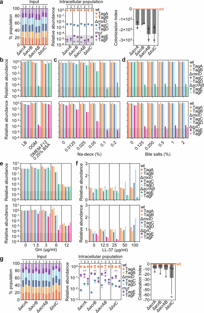
Studying the pathogenesis of human-adapted microorganisms is challenging, since small animal models often fail to recapitulate human physiology. Hence, the comprehensive genetic and regulatory circuits driving the infection process of principal human pathogens such as Shigella flexneri remain to be defined. We combined large-scale Shigella infections of enteroids and colonoids with transposon-directed insertion sequencing and Bayesian statistical modeling to address infection bottlenecks, thereby establishing the comprehensive genome-wide map of Shigella genes required to infect human intestinal epithelium. This revealed the Shigella virulence effectors essential for epithelial cell colonization across geometries and intestinal segments, identified over 100 chromosomal genes involved in the process and uncovered a post-transcriptional mechanism whereby tRNA-modification enzymes and differential codon usage exert global control of a bacterial virulence program. Our findings provide a broadly applicable framework for combining advanced organotypic tissue culture with functional genomics and computational tools to map human-microorganism interactions at scale.


Conflict of interest statement

Competing interests: The authors declare no competing interests.

Figures

Fig. 1
Fig. 1. Scalable human intestinal epithelium Shigella infection model.
a,b, Representative time-lapse series of a BO enteroid infected with wild-type Shigella harboring cytosolic reporter puhpT-GFP (a) or constitutive reporter p-mCherry (b) at MOI 40. See also Supplementary Videos 1 and 2. Cartoon depicts the imaging plane. Experiment repeated twice with imaging of at least ten enteroids per experiment. Lu., lumen. c, Shigella CFU counts (top) and percentage of inoculum (bottom) upon coinfection of BO enteroids with wild-type (WT) and ΔmxiD (noninvasive) strains for 6 h at MOI 40; n = 7 biological replicates pooled from three independent experiments. Data shown as mean ± s.d. Significance determined by two-sided Mann–Whitney U test; ***P < 0.001. d, BO enteroids were infected with a Shigella Tn5 random mutant library containing ~10,000 or ~30,000 mutants. Shown is the number of unique TIS mapped in input (dark gray) or intracellular population output libraries (red). e, Simulations of the number of unique TIS in output samples for bottleneck sizes up to 6,000 bacteria (solid lines) and the measured number of unique TIS (dashed lines). Gray area indicates the plausible range of bottleneck sizes based on the simulations. f, Shigella Tn5 random mutant library 1 (~130,000 mutants) was grown overnight at 30 °C or 37 °C and the mutant abundance compared by TraDIS with respect to the subculture (Methods). Shown is the number of significant advantageous (Adv) (blue; gene presence favors growth) or detrimental (Detr) (orange; gene presence supresses growth) genes; log2FC ≥ 1, FDR ≤ 0.01. g, Venn diagram of Shigella differentially expressed genes in the following comparisons: wild-type, 37 °C versus 30 °C (temperature-regulated genes), wild-type versus ΔvirF 37 °C (VirF-regulated genes) and wild-type versus ΔvirF 30 °C; log2FC ≥ 1, FDR ≤ 0.01. h, Graph showing relative mutant fitness for pINV-located genes (log2FC as noted in Extended Data Fig. 2d; Enr 37 °C versus Enr 30 °C) versus the respective expression changes (RNA-seq; log2FC between wild-type Shigella 37 °C versus 30 °C; Extended Data Fig. 2e). Significant pINV advantageous (dark blue; gene presence favors growth) or detrimental (dark orange; gene presence supresses growth) genes are shown. Two selected chromosomal virF regulator genes, cpxR and fis, also indicated in light orange; log2FC ≥ 1; FDR ≤ 0.01. i, Barcoded competition assay with a consortium comprising three Shigella wild-type (tagA, tagB, tagC), two ΔmxiD (tagD, tagE) and two ΔvirF (tagF, tagG) strains grown overnight at 30 °C or 37 °C. Left, percentage of each strain in the input populations. Right, quantification of relative strain abundances in the Shigella barcoded consortia cultures grown at 30° or 37 °C. Relative abundances normalized to the input inoculum. Data for three independently generated consortia. NS, not significant. j, As in i, but using a consortium comprising three Shigella wild-type (tagA, tagB, tagC), two ΔmxiD (tagD, tagE) and two ΔvirB (tagF, tagG) strains. Significance in i and j determined by two-sided paired t-test between normalized output and normalized input abundances (Methods). *P < 0.05. Panels a and b partially created using BioRender.com. Source data
Fig. 2
Fig. 2. Genome-wide map of the Shigella geneset required to invade the human enteroid infection model.
a, Schematic of the optimized protocol used for the Shigella TraDIS screen in human BO enteroids. b, Shigella M90T chromosome and pINV maps showing the distribution of TIS across the 43 input sub-libraries (gray) versus input sub-library 1 only (blue); ori, origin of replication for the chromosome and pINV; ER, entry region for the pINV. c, Number of unique TIS and total number of sequencing read counts across all input and output sub-libraries. Sub-library 11 was removed in the subsequent analysis due to the low number of sequencing read counts in the input sample. d, Schematic of the ZINB model developed to map Shigella genome-wide colonization factors in the presence of infection bottlenecks. Workflow shows the TraDIS raw dataset used for the sequential fitting of ZINB model parameters. The ZINB model was first applied to TIS in pseudogenes only to fit technical parameters (pairwise normalization factors and the dependence of stochastic mutant loss on input abundance αrgk,in and the fraction of zeros in the output sample fz,r). These fitted parameters were then used in a second ZINB model applied to all ~85,000 TIS to extract gene-wise log2FC values and determine significance. e,f, MA plots showing chromosome-located (e) or pINV-located (f) Shigella gene mutant relative abundances upon infection of enteroids, as quantified by TraDIS. Shown is the log2FC in the output library on the y axis and the log2CPM (average log2-transformed counts per million) on the x axis as detailed in d; each dot represents a gene. CDS, coding sequence; Pseudo, yellow, pseudogene. Panel a partially created using BioRender.com.
Fig. 3
Fig. 3. T3SS effector contributions to Shigella IEC colonization across different cell geometries and intestinal segments.
a, Heat map showing log2FC for T3SS effector gene mutants, as informed by the TraDIS screen in Fig. 2f; **FDR < 0.1; *FDR < 0.2. be, Representative confocal fluorescent microscopy images of a BO enteroid (b), AO enteroid (c), BO colonoid (d) and AO colonoid (e) infected with wild-type Shigella harboring cytosolic reporter puhpT-GFP at MOI 40. Experiments were repeated at least twice with imaging of at least ten enteroids or colonoids per experiment. fi, Shigella CFU counts (left) and percentage of inoculum (right) upon coinfection with wild-type and ΔmxiD (noninvasive) strains for 6 h at MOI 40 of BO enteroids (f, same plots as in c), AO enteroids (g), BO colonoids (h) and AO colonoids (i); n = 7 biological replicates pooled from two to three independent experiments. Data shown as mean ± s.d. Significance determined by two-sided Mann–Whitney U-test; ***P < 0.001. jm, BO and AO enteroids (j) and BO and AO colonoids (l) were infected with a barcoded consortium comprising two wild-type (tagA, tagB), two ΔmxiD (tagC, tagD) and two mutant (tagE, tagF) strains at MOI 40 for 6 h. Shown is the quantification of relative tag abundance, normalized against the corresponding input (Extended Data Fig. 5a,b). Data represent two independently generated consortia for each infection. Significance determined by two-sided paired t-test for each specific mutant between normalized output and normalized input abundances (Methods). *P < 0.05, **P < 0.01, ***P < 0.001. km, Bargraph showing the colonization index for each mutant in BO (dark gray) and AO (light gray) enteroid infections (k, derived from data in j and calculated as 1 – (WT/mutant); n as in j) and BO (dark gray) and AO (light gray) colonoid infections (m, derived from data in l; n as in l). Data shown as mean ± s.d. Wild-type shown as 0. Source data
Fig. 4
Fig. 4. Global regulation of pINV-encoded virulence proteins via tRNA modification and differential codon abundance.
a, Schematic of MnmE/G-dependent tRNA modifications. b, BO enteroids were infected with barcoded consortia comprising two wild-type (tagA, tagB), two ΔmxiD (tagC, tagD) and two mutant (tagE, tagF) strains at MOI 40 for 6 h. Left, quantification of relative tag abundance, normalized against the corresponding input (Extended Data Fig. 6d). Data for three independently generated consortia for each infection. Significance determined by two-sided paired t-test between normalized output and normalized input abundances. **P < 0.01. Right, colonization index for the ΔmnmE and ΔmnmG mutants (derived from data in left panel). Data shown as mean ± s.d. c,d, Volcano plots showing differentially abundant proteins in Shigella ΔmnmE versus wild-type (c) or ΔmnmG versus wild-type (d) strains. Each dot represents a protein. Differentially abundant proteins encoded on pINV shown in pink, on chromosome in black and all nonsignificant differentially abundant proteins in gray. Significance determined by two-sided limma analysis with Benjamini–Hochberg multiple comparison correction; log2FC ≥ 0.5; adjusted P value ≤ 0.01. e,f, Plots showing relative protein levels (log2FC as in c, ΔmnmE versus wild-type) versus AGA codon usage ratios for Arg (e) and GGA codon usage ratios for Gly (f) per protein. Differentially abundant proteins encoded on pINV shown in pink, on chromosome in black, and nonsignificant differentially abundant proteins in gray; log2FC ≥ 0.5; adjusted P value ≤ 0.01. Dark and light green dashed lines specify mean AGA/GGA codon usage ratios for all open reading frames on pINV or chromosome, respectively. gi, ipaA (g), mnmE (h) and mnmG (i) mRNA levels (2–ΔΔCt) upon induction with 0.25 mM IPTG in the indicated strains carrying an inducible ipaA-3xFT plasmid, p-Empty or p-mnmE or p-mnmG, normalized to the corresponding mRNA levels in wild-type Shigella. j, Relative IpaA-3xFT protein levels (0.25 mM IPTG) in wild-type Shigella, ΔmnmE or ΔmnmG strains carrying plasmids as in gi. Quantification by western blot of serially diluted samples with wt/ipaA-3xFT protein levels set as 1. Data shown as mean ± s.d. of three independent experiments (Extended Data Fig. 7a,b). k,l, BO enteroids were infected with barcoded consortia comprising the indicated strains at MOI 40 for 6 h. Shown is the quantification of relative tag abundance, normalized on the corresponding input (Extended Data Fig. 7c,d). Data for two independently generated consortia per infection. Significance determined by two-sided paired t-test between normalized output and normalized input abundances. Only consortia including the two mutant strains carrying the p-Empty or p-mnmE (k) p-mnmG (l) plasmids were used in the analysis. *P < 0.05, **P < 0.01, ***P < 0.001. m,n, mnmE (m) and mnmG (n) mRNA expression (2–ΔΔCt) in the indicated strains, normalized to mnmE (m) and mnmG (n) expression in wild-type Shigella. For g,h,i,m and n, n = 3 biological replicates. Data shown as mean ± s.d. Panel a partially created using BioRender.com. Source data
Extended Data Fig. 1
Extended Data Fig. 1. Characteristics of the basal-out enteroid infection model for early Shigella colonization of human intestinal epithelium.
(Extended Data Fig) Representative image showing a BO enteroid suspension culture as it appears before infection. (b) Shigella CFU counts upon coinfection of BO enteroids with a mix of wt and ΔmxiD (non-invasive) strains for 2 h and 6 h at MOI 40. CFU counts come from 5 biological replicates pooled from 2 independent experiments. Data shown as Mean +/- SD. Significance determined by two-sided Mann-Whitney U-test; **p < 0.01. (c) Shigella CFU counts upon coinfection of Caco-2 cells with a mix of wt and ΔmxiD (non-invasive) strains for 2 h and 6 h at MOI 40. CFU counts come from 7 biological replicates pooled from 2 independent experiments. Data shown as Mean +/- SD. Significance determined by two-sided Mann-Whitney U-test; **p < 0.01; ***p < 0.001. (d) Graph showing the number of generations (left panel) and the generation time (right panel) for Shigella intracellular populations in BO enteroids and Caco-2 cells between 2 and 6 h pi. Number of generations was calculated using CFUs counts from Extended Data Fig. 1b-c. Data shown as Mean +/- SD. Significance determined by two-sided Mann-Whitney U-test; **p < 0.01. (e) Representative time-lapse series of BO enteroids stained with DRAQ7 and treated as following: uninfected (first row); uninfected with addition of saponin after 4 h (second row) and, infected with Shigella wt containing the cytosolic reporter puhpT-GFP at MOI 40 (third row). Experiments were repeated 4 times with imaging of at least 10 enteroids/experiment. Roman numbers represent five different examples of infection foci. Scale bar: 50 µm. Source data
Extended Data Fig. 2
Extended Data Fig. 2. Optimization of experimental design for combining TraDIS with Shigella enteroid infections.
(a) Shigella M90T chromosome and pINV maps showing the TIS distribution across the Tn5 Library #1 (grey). Origin of replication (ori) for chromosome and pINV and entry region (ER) for pINV in magenta. (b–d) MA plots of relative mutant fitness expressed as Log2FC between Enr 30 °C vs Sub (b), Enr 37 °C vs Sub (c) and Enr 37 °C vs Enr 30 °C (d). Each dot represents a gene. Detrimental genes located on pINV shown in dark orange, detrimental genes on the chromosome in light orange, advantageous genes on the pINV in dark blue, and advantageous genes on the chromosome in light blue. Significance determined by two-sided F-test with Benjamini-Hochberg correction for multiple hypothesis testing. Log2FC ≥ 1; FDR ≤ 0.01. (e–g) Volcano plots showing differentially expressed genes in Shigella wt grown at 37 °C vs 30 °C (e), Shigella wt vs ΔvirF mutant grown at 37 °C (f) and Shigella wt vs ΔvirF mutant grown at 30 °C (g). Each dot represents a gene. Differentially expressed genes located on the pINV shown in pink, on the chromosome in black, and all non-significant differentially expressed genes in grey. Significance determined by two-sided F-test with Benjamini-Hochberg correction for multiple hypothesis testing. Log2FC ≥ 1; FDR ≤ 0.01. (h) Plot showing relative mutant fitness for both pINV- and chromosome-located genes on the y-axis (Log2FC as noted in Extended Data Fig. 2d – Enr 37 °C vs Enr 30 °C) versus the respective expression changes on the x-axis (RNA-Seq – Log2FC as noted in Extended Data Fig. 2e for Shigella wt grown at 37 °C vs 30 °C). Significant pINV-encoded advantageous (dark blue) or detrimental (dark orange) and chromosomal advantageous (light blue) or detrimental (light orange) genes are shown. Log2FC ≥ 1; FDR ≤ 0.01. (i) Schematic representation of the tag-containing Shigella chromosomal locus. Primer binding sites for qPCR tag quantification indicated. (j) Standard curves for qPCR detection of tags A-G, using specific primer pairs (see Supplementary Table 3). gDNA from pure monocultures of each of seven Shigella wt tagA-G barcoded strains were diluted (ten-fold dilutions up to 10−6) and used to generate standard curves for each primer pair. R2, slope (Y) and efficiency (E) values indicated in each panel. Source data
Extended Data Fig. 3
Extended Data Fig. 3. Shigella mutant library characteristics and modelling p rameters.
(a) In silico estimation of the number of genes with at least 3 TIS for input sub-libraries with 1,000-3,000 mutants. (b) Pie chart showing expected gene-wise TIS number across input sub-libraries of 40 replicates with 3,000 mutants each, based on the simulation in Extended Data Fig. 3a. 17% of genes had no TIS, ~1,371 ≥ 20 TIS. (c) The Shigella Tn5 mutant library #2 for enteroid infections pooled into 43 sub-libraries. Shown is the number of mutants/sub-library determined by CFU plating. (d) Total Shigella intracellular population sizes upon enteroid infection with sub-libraries in c, determined by CFU plating. No data exist for sub-libraries 4-9. (e) Pie chart showing the gene-wise TIS number across samples of the 42 Shigella Tn5 mutant input sub-libraries. Sub-library 11 removed due to low input sequencing read counts (see Fig. 2c). 14% of genes had no TIS, ~1,219 genes ≥20 TIS. (f) Plate diagram depicting the ZINB model to map Shigella invasion factors (see also Fig. 2d). Technical parameters (normalization nr, dependence of mutant loss on abundance αrgk,in and fraction of zeros in the output sample fz,r) estimated from pseudogenes in grey, gene-fitness scores (logFCg) and disperson (Φg) estimated from all genes in red. (g) PCA plot of TraDIS data from the Shigella enteroid infections. Input-output sub-libraries cluster by replicate. (h) Histogram showing that distribution of read counts of input TIS can be approximated by a negative binomial distribution (red line). Infection bottleneck leads to zero-inflated count distribution in output samples. (i) Fraction of TIS with zero counts in output (and non-zero counts in the respective input) varies between ~25 and ~75% across sub-libraries. (j) Fraction of zero counts in the output depends on the input TIS abundance. Y-axis shows fraction of zero counts, x-axis TIS input abundance normalized by average TIS input abundance. (k) Histogram showing the distribution of z-values approximated by a normal distribution around zero with a standard deviation of 0.9 (see Methods, Supplementary Note 1). (l) The FDR corresponds to the cumulative fraction of z-values above and below the normal distribution (see Methods). Dashed line indicates an FDR cut-off of 0.01. Source data
Extended Data Fig. 4
Extended Data Fig. 4. Contribution of the AcrABTolC efflux pump system to Shigella colonization of enteroids, and resilience to environmental conditions relevant to the infection assay.
(a) BO enteroids were infected with barcoded consortia comprising two Shigella wt (tagA, tagB), two ΔmxiD (tagC, tagD) and two mutant (tagE, tagF) strains at MOI 40 for 6 h in Organoid growth medium (OGM). Mut refers to mutant specified on x-axis. Left panel, percentage of each strain in the input population. Middle panel, relative tag abundance in the intracellular population, normalized against the corresponding input (see Left panel). Right panel, Colonization Index for the indicated mutants (derived from data in the middle panel and calculated as 1-(wt/mut)). Data in right panel shown as Mean +/- SD. Data for two independently generated consortia per infection. Grey shading indicates detection limit. Note that relative abundances of ΔacrA, ΔacrB, ΔacrAB, and ΔtolC mutants fall under the detection limit and are ~1000-fold lower than the non-invasive ΔmxiD strain. (b–f) Barcoded competition assay with a consortium comprising two wt (tagA, tagB), two ΔmxiD (tagC, tagD) and two ΔacrAB or ΔtolC mutant (tagF, tagG) strains grown in different media (LB broth, Organoid growth medium (OGM) or DMEM-F12/0.25%BSA) (b), or LB broth with increasing concentration of Na-deoxycholate (c), Bile salts (d), gentamicin (e), or LL-37 (f). Shown is the relative tag abundance under each condition. Relative tag abundances normalized to the consortia grown in LB. (g) BO enteroids infected with barcoded consortia as in a, but in DMEM-F12/0.25%BSA medium. Data for three independently generated consortia per infection. Significance determined by two-sided paired t-test between normalized output and input abundances (see Methods). **p < 0.01. Data in right panel shown as Mean +/- SD. Interpretation: ΔacrA, ΔacrB, ΔacrAB and ΔtolC mutants were not only sensitive to bile salts/deoxycholate, but also to the OGM medium used for infections (see b-d). Therefore, the low Log2FC value observed for these mutants in the TraDIS screen does not exclusively reflect decreased enteroid colonization, but also generalized sensitivity to multiple infection condition stressors (a–f). Nevertheless, an epithelial colonization defect was validated in the absence of OGM and Na-deoxycholate. Here, the Colonization Indexes for ΔacrA, ΔacrB, ΔacrAB mutants were ~10-fold lower and for ΔtolC mutant ~20-30-fold lower than the wt (g; compare to a). Source data
Extended Data Fig. 5
Extended Data Fig. 5. Input compositions and phenotypic validations for barcoded consortium infections with strains carrying mutations in Shigella T3SS effecto s.
(a, b) BO and AO enteroids (a) and BO and AO colonoids (b) were infected with a mixed barcoded consortium comprising two wt (tagA, tagB), two ΔmxiD (tagC, tagD) and two mutant (tagE, tagF) strains at MOI 40 for 6 h. Mut refers to the specific mutant as indicated on x-axis. Graphs depict the composition of barcoded Shigella consortia used as input inoculums for experiments in Fig. 3j–m. The relative abundance of each tag in the input consortia is plotted as percentage of the total population. Note that no strain is consistently over- or underrepresented. Shown are data for two independently generated consortia for each infection. See Fig. 3j–m for the corresponding relative tag abundances in the intracellular population and Colonization Index scores. (c–f) BO and AO enteroids (ID 22-2jej) (c) and BO and AO colonoids (ID 22-4col) (e) were infected with a mixed barcoded consortium comprising two wt (tagA, tagB), two ΔmxiD (tagC, tagD) and two ΔipaA (tagE, tagF) strains at MOI 40 for 6 h. Shown is the quantification of relative tag abundance, normalized against the corresponding input. For each geometry, infections were executed with four independently generated consortia. Significance determined by two-sided paired t-test for each specific mutant between the normalized output and the normalized input abundances (see Methods). ***p < 0.001. (d–f) Bar graph showing the Colonization Index for each mutant in BO (dark grey) and AO (light grey) enteroid 22-2jej infections (derived from data in c and calculated as 1-(wt/mut); sample size as in c) and BO (dark grey) and AO (light grey) colonoid 22-4col infections (derived from data in e and calculated as 1-(wt/mut); sample size as in e). wt is shown as 0. Data shown as Mean +/- SD. Source data
Extended Data Fig. 6
Extended Data Fig. 6. Enteroid colonization phenotypes for chromosomal Shigella genes, and analysis of codon usage b.
(a–c) BO enteroids infected with barcoded consortia comprising two wt (tagA, tagB), two ΔmxiD (tagC, tagD) and two mutant (tagE, tagF) strains at MOI 40 for 6 h. Mut refers to the mutant specified on x-axis. (a) Percentage of each strain in the input and (b) relative tag abundance in the intracellular population, normalized against the corresponding input. Data for two independent consortia per infection. Significance determined by two-sided paired t-test (see Methods). ns - non-significant; *p < 0.05. (c) Colonization Indexes for the indicated mutants (Derived from b and calculated as 1-(wt/mut)). Data shown as Mean +/- SD. (d) Percentage of each strain in the input for barcoded consortia used for infections in Fig. 4b. (e) Intracellular bacteria in BO enteroids infected with Shigella wt, ΔmxiD, ΔmnmE or ΔmnmG for 3 h at MOI 40. CFU counts from 6 (wt), 5 (ΔmxiD) and 7 (ΔmnmE/ ΔmnmG) biological replicates pooled from 2 independent experiments, normalized to wt. Data shown as Mean +/− SD. Significance determined by two-tailed Mann-Whitney U-test; ns - non-significant; *p < 0.05; **p < 0.01. (f) Venn-diagram of Shigella differentially expressed proteins in ΔmnmE vs wt (MnmE regulated proteins), and ΔmnmG vs wt (MnmG regulated proteins); Log2FC ≥ 0.5, adj_pvalue ≤ 0.01. (g) Linear regression between pINV differentially expressed proteins in ΔmnmE vs wt (MnmE regulated proteins), and ΔmnmG vs wt (MnmG regulated proteins); Log2FC ≥ 0.5, adj_pvalue ≤ 0.01. (h) Codon usage frequency in pINV-located Shigella genes compared to chromosomal genes. Dashed line indicates no enrichment. (i, j) Relative protein levels (Log2FC as in Fig. 4d – ΔmnmG vs wt) versus AGA codon usage ratios for Arg (i) and GGA for Gly (j) per protein. (k–p) Relative protein levels (Log2FC as in Fig. 4c – ΔmnmE vs wt) versus UUA codon usage ratios for Leu (k), CAA for Gln (l), GAA for Glu (m), AAA for Lys (n), AGG for Arg (o) and GGG for Gly (p) per protein. For i-p, differentially expressed proteins on pINV shown in pink, on chromosome in black, and all non-significant differentially expressed proteins in grey. Log2FC ≥ 0.5; adj_pvalue ≤ 0.01. Dark and light green dashed lines specify mean codon usage for all open reading frames on pINV, or chromosome, respectively. Source data
Extended Data Fig. 7
Extended Data Fig. 7. Effects of mnmE and mnmG complementation on IpaA-3xFT protein translation and Shigella fitness during inoculum growth.
(a) Relative IpaA-3xFT protein content in Shigella wt and ΔmnmE strains carrying an inducible pTac-ipaA-3xFT plasmid and the p-Empty or p-mnmE plasmids, determined by quantification of western blots of serially diluted samples. Shown are 3 independent western blots of serially diluted samples for each strain, used for generating standard curves and calculating the relative protein abundance. See Fig. 4j for the corresponding IpaA-3xFT protein content quantification. (b) Relative IpaA-3xFT protein content in Shigella wt and ΔmnmG strains carrying an inducible pTac-ipaA-3xFT plasmid and the p-Empty or p-mnmG plasmids, determined by quantification of western blots of serially diluted samples. Shown are 3 independent western blots of serially diluted samples for each strain, used for generating standard curves and calculating the relative protein abundance. See Fig. 4j for the corresponding IpaA-3xFT protein content quantification. (c) BO enteroids were infected with mixed barcoded consortia comprising the indicated strains at MOI 40 for 6 h, as shown in Fig. 4k. The relative abundance of each tag in the input consortia is plotted as percentage of the total population. Shown are data for two independently generated consortia for each infection. (d) BO enteroids were infected with mixed barcoded consortia comprising the indicated strains at MOI 40 for 6 h, as shown in Fig. 4l. The relative abundance of each tag in the input consortia is plotted as percentage of the total input population. Shown are data for two independently generated consortia for each infection. Source data

References

    1. Schroeder, G. N. & Hilbi, H. Molecular pathogenesis of Shigella spp.: Controlling host cell signaling, invasion, and death by type III secretion. Clin. Microbiol. Rev.21, 134–156 (2008). - PMC - PubMed
    1. Kotloff, K. L., Riddle, M. S., Platts-Mills, J. A., Pavlinac, P. & Zaidi, A. K. M. Shigellosis. Lancet391, 801–812 (2018). - PubMed
    1. Alphonse, N. & Odendall, C. Animal models of shigellosis: a historical overview. Curr. Opin. Immunol.85, 102399 (2023). - PubMed
    1. Marteyn, B., Gazi, A. & Sansonetti, P. Shigella: a model of virulence regulation in vivo. Gut Microbes3, 104–120 (2012). - PMC - PubMed
    1. Bajunaid, W. et al. The T3SS of Shigella: expression, structure, function, and role in vacuole escape. Microorganisms8, 1933 (2020). - PMC - PubMed

MeSH terms

LinkOut - more resources