Review
. 2025 May 28;15(11):1355.
doi: 10.3390/diagnostics15111355.

Non-Coding RNAs in Diagnostic Pathology of High-Grade Central Osteosarcoma

Affiliations
Review

Non-Coding RNAs in Diagnostic Pathology of High-Grade Central Osteosarcoma

Albert Roessner et al. Diagnostics (Basel). .

Abstract

A histological evaluation remains the cornerstone of diagnosing highly malignant osteosarcoma, having demonstrated its efficacy and reliability over several decades. However, despite these advancements, misdiagnoses with severe consequences, including inadequate surgical procedures, continue to occur. Consequently, there is a pressing need to further enhance diagnostic security. Adjunct immunohistochemical approaches have demonstrated significant effectiveness in regard to cancer diagnostics, generally. However, their utility for identifying highly malignant osteosarcoma is limited. Molecular genetic findings have significantly improved the diagnosis of Ewing's sarcoma by identifying specific translocations and have been used to detect specific IDH gene mutations in chondrosarcoma. Nevertheless, molecular genetic alterations in highly malignant osteosarcoma exhibit a high degree of complexity, thereby limiting their diagnostic utility. Given that only 1-2% of the human genome comprises protein-coding sequences, the growing number of non-coding regulatory RNAs, which are increasingly being elucidated, has garnered substantial attention in the field of clinical cancer diagnostics. Over the past several years, patterns of altered non-coding RNA expression have been identified that facilitate the distinction between benign and malignant tumors in various organs. In the field of bone tumors, the experience of this approach has been limited thus far. The divergent expression of microRNAs has demonstrated utility for differentiating osteosarcoma from osteoblastoma and discriminating between osteosarcoma and giant-cell tumors of bone and fibrous dysplasia. However, the application of non-coding RNA expression patterns for the differential diagnosis of osteosarcoma is still in its preliminary stages. This review provides an overview of the current status of non-coding RNAs in osteosarcoma diagnostics, in conjunction with a histological evaluation. The potential of this approach is discussed comprehensively.

Keywords: differential diagnosis; highly malignant osteosarcoma; non-coding RNAs.


Conflict of interest statement

The authors declare no conflict of interest.

Figures

Figure 5
Figure 5
There is no correlation between the number of protein-coding genes and the developmental complexity of a species. Instead, there is a negative correlation between the percentage of the genome occupied by protein-coding sequences and developmental complexity [67] (Created with BioRender).
Figure 1
Figure 1
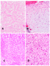
(A) Typical osteoblastic osteosarcoma, with ample osteoid formation appearing as broad homogenously stained trabeculae (H&E ×200). (B) Chondroblastic osteosarcoma, with obvious cartilage nature of the sarcoma tissue (H&E ×200) (C) Fibroblastic osteosarcoma, with typical fibroblast-like spindle cells. This pattern resembles connective tissue. Its malignant nature is revealed by nuclear pleomorphism and mitoses (H&E ×200). (D) Giant cell–rich osteosarcoma, containing abundant osteoclast-like giant cells, with multiple regularly formed nuclei (H&E ×200). (All histological images presented in this manuscript originate from the senior author’s personal archive).
Figure 2
Figure 2
(A) Aggressive osteoblastoma, with atypical cellular nuclei. The pronounced vascularization is typical of osteoblastoma (H&E ×200). (B) Highly malignant osteoblastic osteosarcoma, with a high level of nuclear pleomorphism and typical osteoid formation, indicted by the tumor cells appearing as broad trabeculae (H&E ×200). (C) Low-grade intramedullary osteosarcoma. In this typical case, the bone structure is very highly differentiated and barely discernable from reactive bone formation (H&E ×200). (D) Heterotopic ossification mimicking osteosarcoma. In particular, the immature bone trabeculae are very similar to osteosarcoma (H&E ×200).
Figure 3
Figure 3
(A) Osteoblastic osteosarcoma before preoperative chemotherapy. Viable malignant tumor cells, lacking any signs of regression or necrosis. Note the well-stained nuclei of the viable tumor cells (H&E ×100). (B) Completely devitalized necrotic area of the former osteoblastic osteosarcoma, with no viable tumor cells left after preoperative chemotherapy. In sharp contrast to the preoperative tissue, no stained nuclei can be observed (H&E ×100). Inset: high-power view of completely devitalized former osteoblastic tumor tissue and remnants of an osteoid. Not one single viable tumor cell is left (H&E ×400).
Figure 4
Figure 4
(A) Typical histological appearance of primary osteoblastic osteosarcoma. The patient did not develop lung metastases. The pleomorphic tumor cells reveal its malignant nature (H&E ×200). (B) Primary osteoblastic osteosarcoma, with lung metastasis, at the time of diagnosis (H&E ×200). The non-metastasizing and metastasizing malignant tumors appear histologically indistinguishable.

References

    1. Yoshida A. Osteosarcoma: Old and New Challenges. Surg. Pathol. Clin. 2021;14:567–583. doi: 10.1016/j.path.2021.06.003. - DOI - PubMed
    1. Savage S.A., Mirabello L. Using epidemiology and genomics to understand osteosarcoma etiology. Sarcoma. 2011;2011:14–16. doi: 10.1155/2011/548151. - DOI - PMC - PubMed
    1. Nagano A., Matsumoto S., Kawai A., Okuma T., Hiraga H., Matsumoto Y., Nishida Y., Yonemoto T., Hosaka M., Takahashi M., et al. Osteosarcoma in patients over 50 years of age: Multi-institutional retrospective analysis of 104 patients. J. Orthop. Sci. 2020;25:319–323. doi: 10.1016/j.jos.2019.04.008. - DOI - PubMed
    1. Baumhoer D., Brunner P., Eppenberger-Castori S., Smida J., Nathrath M., Jundt G. Osteosarcomas of the jaws differ from their peripheral counterparts and require a distinct treatment approach. Experiences from the DOESAK Registry. Oral Oncol. 2014;50:147–153. doi: 10.1016/j.oraloncology.2013.10.017. - DOI - PubMed
    1. Robinson M.J., Davis E.J. Neoadjuvant Chemotherapy for Adults with Osteogenic Sarcoma. Curr. Treat. Options Oncol. 2024;25:1366–1373. doi: 10.1007/s11864-024-01269-2. - DOI - PMC - PubMed

LinkOut - more resources