

Where Reflectance Confocal Microscopy Provides the Greatest Benefit for Diagnosing Skin Cancers: The Experience of the National Cancer Institute of Naples
- PMID: 40507227
- PMCID: PMC12153731
- DOI: 10.3390/cancers17111745
Where Reflectance Confocal Microscopy Provides the Greatest Benefit for Diagnosing Skin Cancers: The Experience of the National Cancer Institute of Naples
Abstract
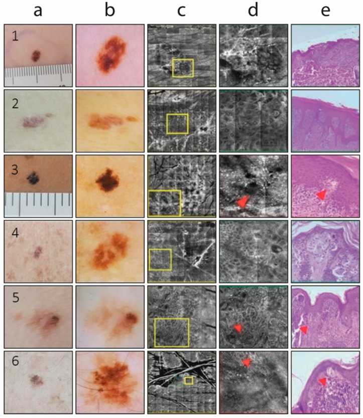
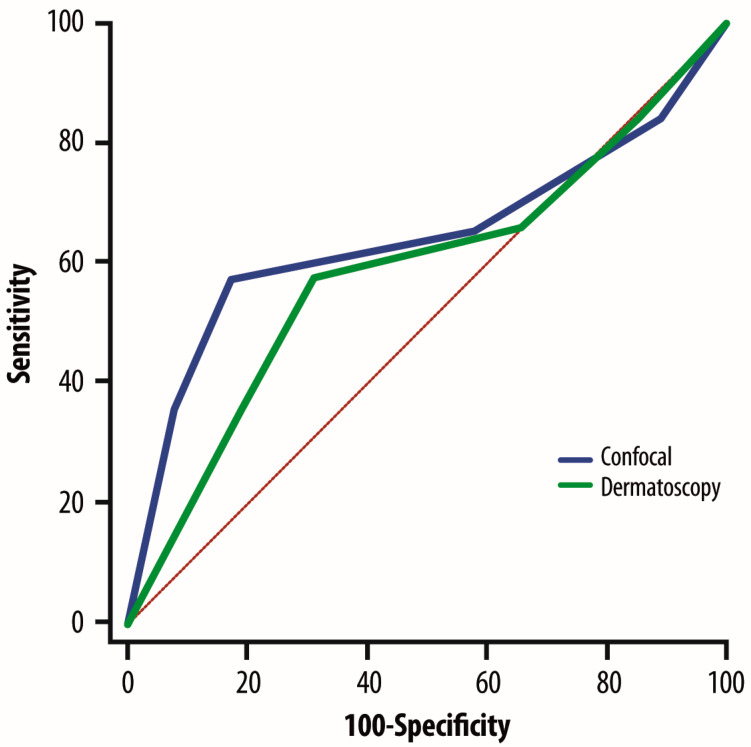
Background: Although complete excision of suspicious melanocytic lesions is mandatory, it carries the risk of unnecessary scarring on one hand and inadequate treatment of misdiagnosed lesions on the other. Objectives: We evaluated the sensitivity, specificity, and predictive value of reflectance confocal microscopy (RCM) in diagnosing pigmented lesions with clinically ambiguous features-the so-called "gray zone" -and compared its performance with the more established technique of epiluminescence microscopy (ELM). Results: Between 2019 and 2020, a total of 2282 melanocytic lesions were assessed using both ELM and RCM. Histopathological diagnosis aligned with the ELM risk classification in 91.6% of melanocytic lesions, specifically in 92.0% of very-high-risk lesions, 88.5% of high-risk lesions, 66.3% of medium-risk lesions, 96.3% of low-risk lesions, and 98.0% of very low-risk lesions. Similarly, histopathological diagnosis of these lesions corresponded with the RCM risk assessment in 91.2% of cases, including 90.9% of very-high-risk lesions, 84.4% of high-risk lesions, 93.1% of medium-risk lesions, 90.5% of low-risk lesions, and 96.2% of very low-risk lesions. Conclusions: Although ELM is a valuable tool for increasing the efficacy of clinical diagnosis, its reliability decreases for a group of lesions that appear suspicious during clinical skin examination. RCM, as a newer technique, appears to improve malignancy detection in suspicious melanocytic lesions without requiring excision; its sensitivity and specificity remain high even in lesions classified by ELM as posing a medium risk of malignancy.
Keywords: epiluminescence microscopy; melanocytic lesions; melanoma; reflectance confocal microscopy.
Conflict of interest statement
All the authors declare no competing interests except for P.A.A. He has/had a consultant/advisory role for Bristol Myers Squibb, Roche-Genentech, Merck Sharp and Dohme, Novartis, Merck Serono, Pierre-Fabre, AstraZeneca, Sun Pharma, Sanofi, Idera, Sandoz, Immunocore, 4SC, Italfarmaco, Nektar, Boehringer-Ingelheim, Eisai, Regeneron, Daiichi Sankyo, Pfizer, Oncosec, Nouscom, Lunaphore, Seagen, iTeos, Medicenna, Bio-Al Health, ValoTX, and Replimmune. He also received research funding from Bristol Myers Squibb, Roche-Genentech, Pfizer, and Sanofi, as well as travel support from Pfizer. The funders of this study had no role in the design of the study; in the collection, analyses, or interpretation of data; in the writing of the manuscript, or in the decision to publish the results.
Figures
Similar articles
-
Role of In Vivo Reflectance Confocal Microscopy in the Analysis of Melanocytic Lesions.Acta Dermatovenerol Croat. 2018 Apr;26(1):64-67. Acta Dermatovenerol Croat. 2018. PMID: 29782304 Review.
-
Reflectance confocal microscopy for diagnosing cutaneous melanoma in adults.Cochrane Database Syst Rev. 2018 Dec 4;12(12):CD013190. doi: 10.1002/14651858.CD013190. Cochrane Database Syst Rev. 2018. PMID: 30521681 Free PMC article.
-
Reflectance confocal microscopy for diagnosing keratinocyte skin cancers in adults.Cochrane Database Syst Rev. 2018 Dec 4;12(12):CD013191. doi: 10.1002/14651858.CD013191. Cochrane Database Syst Rev. 2018. PMID: 30521687 Free PMC article.
-
Epiluminescence microscopy as a useful approach in the early diagnosis of cutaneous malignant melanoma.Melanoma Res. 1998 Dec;8(6):529-37. doi: 10.1097/00008390-199812000-00008. Melanoma Res. 1998. PMID: 9918415
-
Reflectance confocal microscopy analysis of equivocal melanocytic lesions with severe regression.Skin Res Technol. 2018 Feb;24(1):9-15. doi: 10.1111/srt.12382. Epub 2017 May 21. Skin Res Technol. 2018. PMID: 28543606
References
-
- Gershenwald J.E., Scolyer R.A., Hess K.R., Sondak V.K., Long G.V., Ross M.I., Lazar A.J., Faries M.B., Kirkwood J.M., McArthur G.A., et al. Melanoma staging: Evidence-based changes in the American Joint Committee on Cancer eighth edition cancer staging manual. CA Cancer J. Clin. 2017;67:472–492. doi: 10.3322/caac.21409. - DOI - PMC - PubMed
-
- Garbe C., Amaral T., Peris K., Hauschild A., Arenberger P., Basset-Seguin N., Bastholt L., Bataille V., Brochez L., del Marmol V., et al. European consensus-based interdisciplinary guideline for melanoma. Part 1: Diagnostics—Update 2024. Eur. J. Cancer. 2025;215:115152. doi: 10.1016/j.ejca.2024.115152. - DOI - PubMed
-
- Dinnes J., Deeks J.J., Saleh D., Chuchu N., Bayliss S.E., Patel L., Davenport C., Takwoingi Y., Godfrey K., Matin R.N., et al. Reflectance confocal microscopy for diagnosing cutaneous melanoma in adults. Cochrane Database Syst. Rev. 2018;12:CD013190. doi: 10.1002/14651858.CD013190. - DOI - PMC - PubMed
LinkOut - more resources
Full Text Sources
