

Fatty Pancreas: Its Potential as a Risk Factor for Pancreatic Cancer and Clinical Implications
- PMID: 40507246
- PMCID: PMC12153785
- DOI: 10.3390/cancers17111765
Fatty Pancreas: Its Potential as a Risk Factor for Pancreatic Cancer and Clinical Implications
Abstract
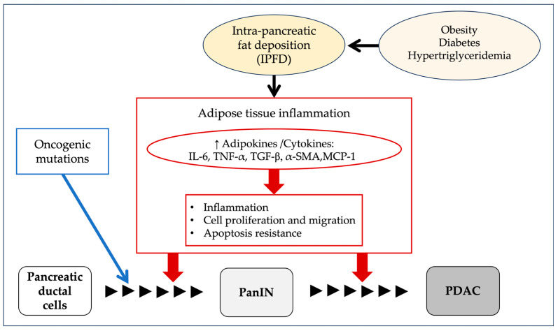
With the increasing use of imaging modalities such as ultrasonography, computed tomography, and magnetic resonance imaging, incidental findings of pancreatic abnormalities, including pancreatic cysts and fatty pancreas (FP), have become more common. FP, also referred to as pancreatic steatosis, intra-pancreatic fat deposition, or fatty pancreas disease, is characterized by the accumulation of fat in the pancreas. Although FP has been associated with metabolic syndromes such as obesity and diabetes, its clinical significance remains unclear. Recent evidence suggests that FP may play a role in pancreatic carcinogenesis. Metabolic disorders, including obesity, insulin resistance, and diabetes, have been implicated in the development of FP. Additionally, FP has been linked to an increased risk of pancreatic ductal adenocarcinoma (PDAC), possibly due to chronic inflammation, lipotoxicity, and an altered pancreatic microenvironment. While early detection of PDAC remains challenging, surveillance strategies for high-risk individuals, such as those with pancreatic cysts, new-onset diabetes, or a genetic predisposition, may be crucial. In this context, FP may be incorporated into this surveillance of high-risk individuals. Some pharmacological interventions, including glucagon-like peptide-1 (GLP-1) receptor agonists and sodium-glucose cotransporter-2 (SGLT2) inhibitors, have shown potential in reducing pancreatic fat accumulation, although further studies are needed to confirm their efficacy.
Keywords: fatty pancreas; intra-pancreatic fat deposition (IPFD); pancreatic ductal adenocarcinoma (PDAC).
Conflict of interest statement
The authors declare no conflicts of interest.
Figures

Similar articles
-
The prevalence and clinical implications of pancreatic fat accumulation identified during a medical check-up.Medicine (Baltimore). 2021 Oct 15;100(41):e27487. doi: 10.1097/MD.0000000000027487. Medicine (Baltimore). 2021. PMID: 34731128 Free PMC article.
-
CT Density in the Pancreas is a Promising Imaging Predictor for Pancreatic Ductal Adenocarcinoma.Ann Surg Oncol. 2017 Sep;24(9):2762-2769. doi: 10.1245/s10434-017-5914-3. Epub 2017 Jun 20. Ann Surg Oncol. 2017. PMID: 28634666
-
Intrapancreatic fat deposition is unrelated to liver steatosis in metabolic dysfunction-associated steatotic liver disease.JHEP Rep. 2024 Jan 1;6(3):100998. doi: 10.1016/j.jhepr.2023.100998. eCollection 2024 Mar. JHEP Rep. 2024. PMID: 38379586 Free PMC article.
-
Association between intra-pancreatic fat deposition and diseases of the exocrine pancreas: A narrative review.World J Gastroenterol. 2025 Jan 14;31(2):101180. doi: 10.3748/wjg.v31.i2.101180. World J Gastroenterol. 2025. PMID: 39811515 Free PMC article. Review.
-
Fatty Pancreas-Centered Metabolic Basis of Pancreatic Adenocarcinoma: From Obesity, Diabetes and Pancreatitis to Oncogenesis.Biomedicines. 2022 Mar 17;10(3):692. doi: 10.3390/biomedicines10030692. Biomedicines. 2022. PMID: 35327494 Free PMC article. Review.
References
-
- Mizuno S., Isayama H., Nakai Y., Yoshikawa T., Ishigaki K., Matsubara S., Yamamoto N., Ijichi H., Tateishi K., Tada M., et al. Prevalence of Pancreatic Cystic Lesions Is Associated With Diabetes Mellitus and Obesity: An Analysis of 5296 Individuals Who Underwent a Preventive Medical Examination. Pancreas. 2017;46:801–805. doi: 10.1097/MPA.0000000000000833. - DOI - PubMed
Publication types
LinkOut - more resources
Full Text Sources
