

Radiation Dose Reduction in Cancer Imaging with New-Model CT Scanners: A Quality of Care Evaluation
- PMID: 40507296
- PMCID: PMC12153870
- DOI: 10.3390/cancers17111815
Radiation Dose Reduction in Cancer Imaging with New-Model CT Scanners: A Quality of Care Evaluation
Abstract
Background/objectives: The primary aim of this study was to evaluate whether the replacement of roughly one-decade-old computed tomography (CT) scanners with new-model CT scanners were associated with an additional reduction in the radiation dose delivered to oncological patients, in a radiological setting where the optimization of protocols had already reached very low radiation doses. An exploratory secondary objective was to evaluate the potential differences in the objective image quality between the CT scans obtained before and after the installation of the new-generation CT scanners.
Methods: Chest and abdominal CT examinations conducted for oncologic purposes were retrospectively selected from two time periods-prior to scanner replacement (2022) and following an upgrade (2024)-after five CT systems in our radiology department were replaced. We extracted and compared the CT dose index (CTDI) and dose length product (DLP) for each CT phase. For the objective image quality evaluation, we calculated the signal-to-noise ratio (SNR) and the contrast-to-noise ratio (CNR) at the center of the liver and the aorta. An appropriate statistical analysis was performed and a p-value < 0.05 was considered significant.
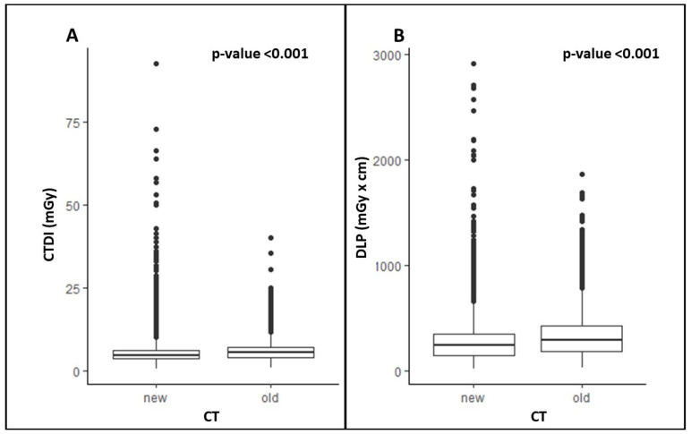
Results: We included 14,601 CT acquisitions, of which 9013 (61.7%) were performed before and 5588 (38.3%) after the replacement of the CT scanners. There were significantly lower values for the CTDI and DLP with the new CT scanners compared to the old ones. The CTDI with the new CT scanners was significantly lower in all phases (p-value = 0.002 for unenhanced phase, and p < 0.001 for arterial, portal venous, and delayed phases). The DLP using the new CT scanners was significantly lower in the arterial, portal venous, and delayed phases (p < 0.001), and it was not significantly different in the unenhanced phase (p = 0.36). There was no significant difference in the SNR at the liver level (p = 0.72) or at the aorta level (p = 0.51). There was no significant difference in the CNR at the liver level (p = 0.24), whereas the CNR was higher with the new CT scanners at the aorta level (p = 0.03).
Conclusions: The transition to new-model CT scanners resulted in a significant reduction in the radiation dose delivered by chest and abdomen CT scans, without compromising the objective image quality.
Keywords: computed tomography; computed tomography dose index; dose length product; objective image quality; radiation dose; replacement.
Conflict of interest statement
The authors declare no conflicts of interest.
Figures


Similar articles
-
Body CT examinations in oncologic patients: the impact of subspecialty radiology on radiation exposure in the clinical practice. A quality care study.Radiol Med. 2024 Mar;129(3):429-438. doi: 10.1007/s11547-024-01790-2. Epub 2024 Feb 11. Radiol Med. 2024. PMID: 38341817 Free PMC article.
-
Dual-energy, standard and low-kVp contrast-enhanced CT-cholangiography: a comparative analysis of image quality and radiation exposure.Eur J Radiol. 2012 Jul;81(7):1405-12. doi: 10.1016/j.ejrad.2011.03.030. Epub 2011 Apr 2. Eur J Radiol. 2012. PMID: 21458939
-
Ultra-low-dose chest CT in adult patients with cystic fibrosis using a third-generation dual-source CT scanner.Radiol Med. 2021 Apr;126(4):544-552. doi: 10.1007/s11547-020-01304-w. Epub 2020 Nov 16. Radiol Med. 2021. PMID: 33200307
-
Evaluation of Radiation Dose and Image Quality in Clinical Routine Protocols from Three Different CT Scanners.J Imaging. 2025 Feb 25;11(3):70. doi: 10.3390/jimaging11030070. J Imaging. 2025. PMID: 40137182 Free PMC article.
-
Image quality of mean temporal arterial and mean temporal portal venous phase images calculated from low dose dynamic volume perfusion CT datasets in patients with hepatocellular carcinoma and pancreatic cancer.Eur J Radiol. 2016 Nov;85(11):2104-2110. doi: 10.1016/j.ejrad.2016.09.024. Epub 2016 Sep 25. Eur J Radiol. 2016. PMID: 27776665
References
LinkOut - more resources
Full Text Sources
