. 2025 May 22;26(11):4990.
doi: 10.3390/ijms26114990.

RANKL Drives Bone Metastasis in Mammary Cancer: Protective Effects of Anti-Resorptive Treatments

Affiliations

RANKL Drives Bone Metastasis in Mammary Cancer: Protective Effects of Anti-Resorptive Treatments

Evi Gkikopoulou et al. Int J Mol Sci. .

Abstract

Receptor activator of nuclear factor-κB ligand (RANKL) is essential for osteoclast formation and bone resorption, in osteolytic conditions such as osteoporosis and bone metastases. However, its role in metastasis progression remains incompletely understood. Herein, we examined whether the overexpression of human RANKL in transgenic mice (TgRANKL) affects their susceptibility to breast cancer bone metastasis compared to their wild-type (WT) littermates. Bone metastasis was induced by injecting EO771 mouse mammary adenocarcinoma cells into the caudal artery of syngeneic WT and TgRANKL mice. RANKL overexpression led to an earlier onset and increased burden of bone metastasis in EO771-bearing TgRANKL mice compared to WT mice. It also exacerbated the bone destruction caused by metastasis-associated osteolysis. The prophylactic inhibition of RANKL activity with denosumab, a monoclonal antibody targeting human RANKL, prevented osteolysis and significantly reduced the incidence and progression of bone metastases in TgRANKL mice. However, the therapeutic denosumab treatment had no effect on metastasis incidence or tumor burden, although it alleviated osteolysis. The treatment with zoledronic acid, an anti-resorptive agent inhibiting osteoclast activity, yielded results similar to those of denosumab. These findings emphasize the significance of initiating early treatment with anti-resorptive agents such as denosumab or zoledronic acid to reduce the risk of bone metastasis in patients at high risk.

Keywords: RANKL; bone metastasis; breast cancer; mouse models; pre-clinical models.


Conflict of interest statement

The authors declare no conflicts of interest.

Figures
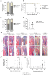
Figure 1
Figure 1
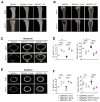
Earlier onset of mammary-cancer-driven bone metastasis and increased metastatic burden in TgRANKL mice. (A) Percentage and absolute number of WT+EO771 and TgRANKL+EO771 mice with metastasis-driven hindlimb paralysis at 21 dpi (n = 10–12/group). (B) Onset kinetics of hindlimb paralysis in mice (n = 6–10/group). Log-rank (Mantel-Cox) test was performed for the statistical analysis. (C) Percentage and absolute number of mice with post-mortem-confirmed ovarian metastasis and (D) mean ovarian tumor weight per mouse. Data are shown as mean values ± SD. Student’s t-test was performed for the statistical analysis (n = 10–12/group). (E) Representative histological images of femurs and tibiae stained with hematoxylin and eosin at the indicated dpi. Dashed yellow lines indicate the metastatic area within the bone, while dotted lines indicate the extraskeletal metastatic area. Scale bar, 150 μm. Quantification of (F) metastatic area/bone area and (G) extraskeletal metastatic area based in histological images of whole hindlimbs from WT+EO771 and TgRANKL+EO771 mice at the indicated dpi. Data are shown as mean values ± SD. Student’s t-test was performed for the statistical analysis between WT and TgRANKL mice at various dpi (n = 5–12/group), * p < 0.05, ** p < 0.001.
Figure 2
Figure 2
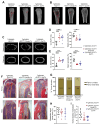
Enhanced mammary-cancer-induced osteolysis in TgRANKL mice. (A,B) Representative micro-CT 3D reconstructed longitudinal images of distal femurs from naïve and EO771-injected WT and TgRANKL mice at 21 dpi. Red lines highlight osteolytic areas. Scale bar, 1 mm. Representative 2D reconstructed cross-section images of the cortical bone at metaphysis (C) and diaphysis (E) with quantification (DF) of cortical bone volume fraction (Ct.BV/TV, %) and total open porosity (%) values in femurs from each study group (n = 5–6/group). Scale bar, 500 μm. A two-way analysis of variance and Tukey’s post hoc test were performed for the statistical analysis; ** p < 0.01, *** p < 0.001, **** p < 0.0001.
Figure 3
Figure 3
Prophylactic administration of denosumab or zoledronic acid prevents mammary-cancer-induced bone metastasis in TgRANKL mice. (A) Schematic representation of the EO771 bone metastasis model with denosumab (Dmab) and zoledronic acid (Zol) treatment strategies. All mice were euthanized at 21 dpi of cancer cells. (B) Relative diameter of the tibiae measured by caliper at the endpoint (n = 6–9/group). (C) Representative photos of in vivo bioluminescence imaging of TgRANKL+EO771, TgRANKL+EO771/Dmab, and TgRANKL+EO771/Zol mice at 19 dpi. Blue arrow indicates bone metastatic signal, while pink arrow indicates ovarian metastasis. (D) Percentage and absolute number of mice with post-mortem-confirmed ovarian metastasis and (E) mean ovarian tumor weight/mouse. (F) Representative images of femurs and tibiae stained with hematoxylin and eosin at 21 dpi. Dashed yellow lines indicate the metastatic area within the bone, while dotted lines indicate the extraskeletal metastatic areas. Scale bar, 150 μm. (G) Percentage and absolute number of mice with histologically verified metastatic tumors in femurs and tibiae. (H) Metastatic area/bone area in hindlimbs and (I) extraskeletal metastatic area (n = 11–15/group). Data are shown as mean values ± SD. A one-way ANOVA and Dunnett’s post hoc test were performed for the statistical analysis, ** p < 0.01, *** p < 0.001.
Figure 4
Figure 4
Prophylactic administration of denosumab or zoledronic acid attenuates osteolysis. (A,B) Representative micro-CT 3D reconstructed longitudinal images of distal femurs from naïve and EO771-injected TgRANKL mice, with or without prophylactic treatment with denosumab (Dmab) or zoledronic acid (Zol). Red lines highlight osteolytic areas. Scale bar, 1 mm. Representative 2D reconstructed cross-section images of the cortical bone at metaphysis (C) and diaphysis (E) with quantification (DF) of the cortical bone volume fraction (Ct.BV/TV, %) and total open porosity (%) in femurs from each group (n = 4–10/group). Scale bar, 500 μm. Data are shown as mean values ± SD. A two-way ANOVA and Dunnett’s post hoc test were performed for the statistical analysis of more than two groups; * p < 0.05, ** p < 0.01, *** p < 0.001, **** p < 0.0001.
Figure 5
Figure 5
Therapeutic administration of denosumab or zoledronic acid prevents osteolysis but does not impact bone metastasis. (A,B) Representative micro-CT 3D reconstructed longitudinal images of distal femurs from naïve and EO771-injected TgRANKL mice, with or without therapeutic treatment with denosumab (Dmab) or zoledronic acid (Zol). Red lines highlight osteolytic areas. Scale bar, 1 mm. (C) Representative 2D reconstructed cross-section images of the cortical bone in the metaphysis with quantification (D,E) of the cortical bone volume fraction (Ct.BV/TV, %) and total open porosity (%) in femurs from each group (n = 6–10/group). Scale bar, 500 μm. (F) Representative images of femurs and tibiae stained with hematoxylin and eosin at 21 dpi. Dashed yellow lines indicate the metastatic area within the bone, while dotted lines indicate the extraskeletal metastatic areas. Scale bar, 150 μm. (G) Percentage and absolute number of each experimental group with histologically verified metastatic tumors in femurs and tibiae. (H) Metastatic area/bone area in hindlimbs and (I) extraskeletal metastatic area (n = 6–11/group). Data are shown as mean values ± SD. A one-way ANOVA and Dunnett’s post hoc test were performed for the statistical analysis of more than two groups; ** p < 0.01, *** p < 0.001, **** p < 0.0001.

References

    1. Bray F., Laversanne M., Sung H., Ferlay J., Siegel R.L., Soerjomataram I., Jemal A. Global Cancer Statistics 2022: GLOBOCAN Estimates of Incidence and Mortality Worldwide for 36 Cancers in 185 Countries. CA Cancer J. Clin. 2024;74:229–263. doi: 10.3322/caac.21834. - DOI - PubMed
    1. Huang J.-F., Shen J., Li X., Rengan R., Silvestris N., Wang M., Derosa L., Zheng X., Belli A., Zhang X.-L., et al. Incidence of Patients with Bone Metastases at Diagnosis of Solid Tumors in Adults: A Large Population-Based Study. Ann. Transl. Med. 2020;8:482. doi: 10.21037/atm.2020.03.55. - DOI - PMC - PubMed
    1. Xiong Z., Deng G., Huang X., Li X., Xie X., Wang J., Shuang Z., Wang X. Bone Metastasis Pattern in Initial Metastatic Breast Cancer: A Population-Based Study. Cancer Manag. Res. 2018;10:287–295. doi: 10.2147/CMAR.S155524. - DOI - PMC - PubMed
    1. Coleman R.E., Brown J., Holen I. Abeloff’s Clinical Oncology. Elsevier; Amsterdam, The Netherlands: 2020. Bone Metastases; pp. 809–830.e3.
    1. Jones D.H., Nakashima T., Sanchez O.H., Kozieradzki I., Komarova S.V., Sarosi I., Morony S., Rubin E., Sarao R., Hojilla C.V., et al. Regulation of Cancer Cell Migration and Bone Metastasis by RANKL. Nature. 2006;440:692–696. doi: 10.1038/nature04524. - DOI - PubMed

MeSH terms

LinkOut - more resources