

Factors Contributing to Resistance to Ischemia-Reperfusion Injury in Olfactory Mitral Cells
- PMID: 40507890
- PMCID: PMC12155091
- DOI: 10.3390/ijms26115079
Factors Contributing to Resistance to Ischemia-Reperfusion Injury in Olfactory Mitral Cells
Abstract
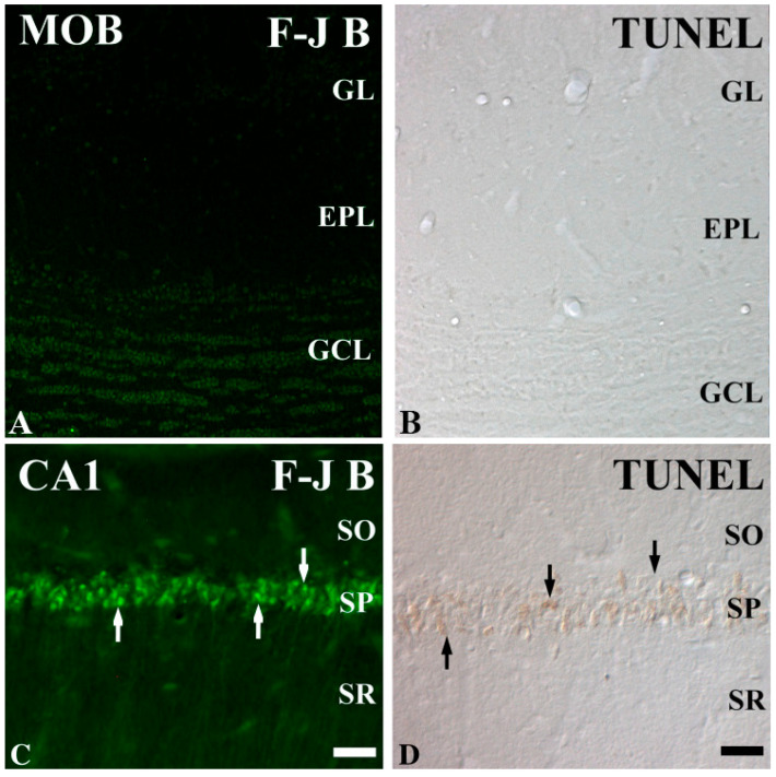
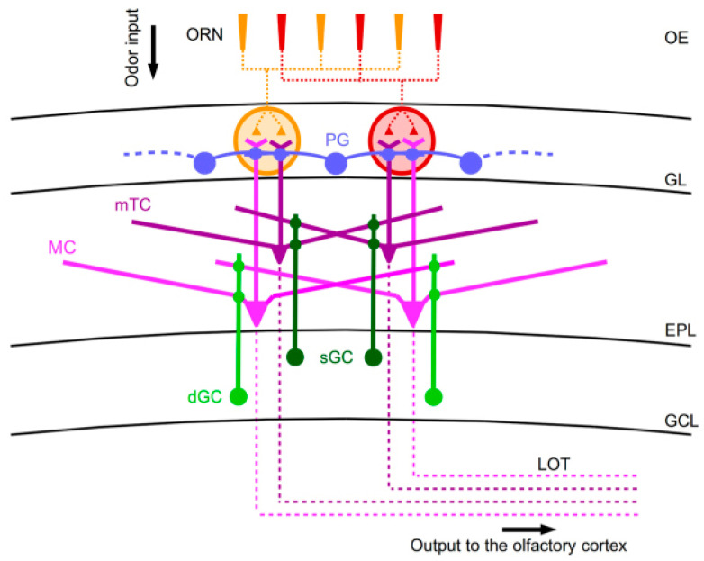
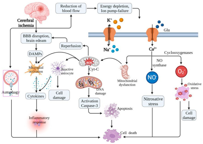
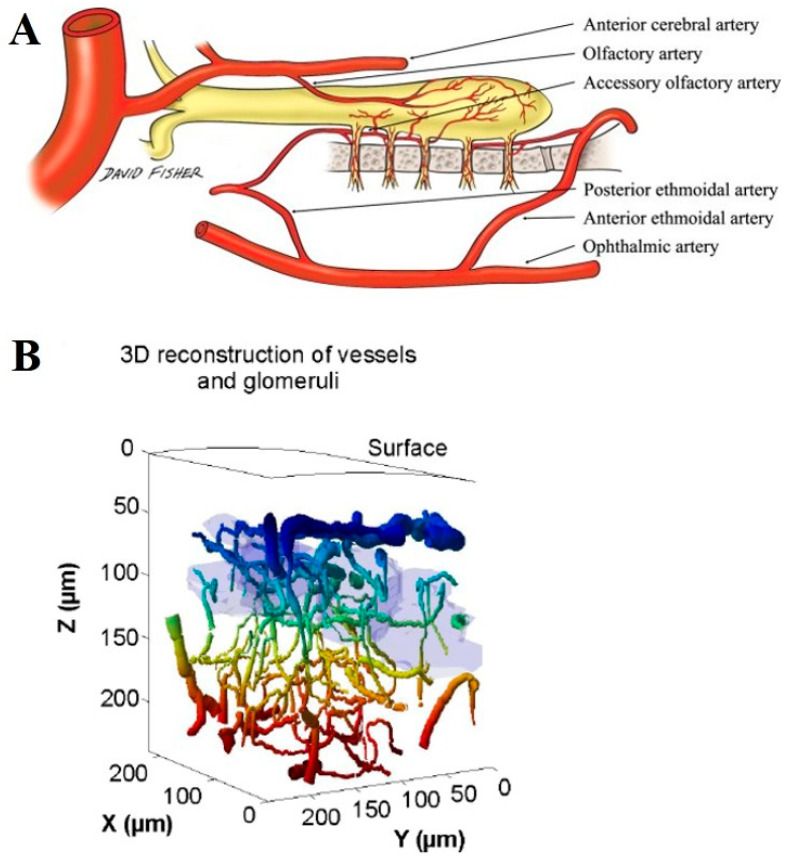
Brain ischemia-reperfusion (IR) injury is a critical pathological process that leads to extensive neuronal death, with hippocampal pyramidal cells, particularly those in the cornu Ammonis 1 (CA1) subfield, being highly vulnerable. Until now, human olfactory mitral cell resistance to IR injury has not been directly studied, but olfactory dysfunction in humans is frequently reported in systemic vascular conditions such as ischemic heart failure and may serve as an early clinical marker of neurological or cardiovascular disease. Mitral cells, the principal neurons of the olfactory bulb (OB), exhibit remarkable resistance to IR injury, suggesting the presence of unique molecular adaptations that support their survival under ischemic stress. Several factors may contribute to the resilience of mitral cells. They have a lower susceptibility to excitotoxicity, mitigating the harmful effects of excessive glutamate signaling. Additionally, they maintain efficient calcium homeostasis, preventing calcium overload-a major trigger for cell death in vulnerable neurons. Mitral cells may also express high baseline levels of antioxidant enzymes and their activities, counteracting oxidative stress. Their robust mitochondrial function enhances energy production and reduces susceptibility to metabolic failure. Furthermore, neuroprotective signaling pathways, including phosphatidylinositol-3-kinase (PI3K)/Akt, mitogen-activated protein kinase/extracellular signal-regulated kinase (MAPK/ERK), and nuclear factor erythroid-2-related factor 2 (Nrf2)-mediated antioxidative responses, further bolster their resistance. In addition to these intrinsic mechanisms, the unique microvascular architecture and metabolic support within the olfactory bulb provide an extra layer of protection. By comparing mitral cells to ischemia-sensitive neurons, key vulnerabilities-such as oxidative stress, excitotoxicity, calcium dysregulation, and mitochondrial dysfunction-can be identified and potentially mitigated in other brain regions. Understanding these molecular determinants of neuronal survival may offer valuable insights for developing novel neuroprotective strategies to combat IR injury in highly vulnerable areas, such as the hippocampus and cortex.
Keywords: antioxidant enzymes; excitotoxicity; metabolic failure; microvascular architecture; neuronal survival; neuroprotective signaling pathways.
Conflict of interest statement
The authors declare no conflicts of interest.
Figures
Similar articles
-
Akt / GSK3β / Nrf2 / HO-1 pathway activation by flurbiprofen protects the hippocampal neurons in a rat model of glutamate excitotoxicity.Neuropharmacology. 2021 Sep 15;196:108654. doi: 10.1016/j.neuropharm.2021.108654. Epub 2021 Jun 11. Neuropharmacology. 2021. PMID: 34119518
-
Meldonium, as a potential neuroprotective agent, promotes neuronal survival by protecting mitochondria in cerebral ischemia-reperfusion injury.J Transl Med. 2024 Aug 15;22(1):771. doi: 10.1186/s12967-024-05222-7. J Transl Med. 2024. PMID: 39148053 Free PMC article.
-
Kirenol alleviates cerebral ischemia-reperfusion injury by reducing oxidative stress and ameliorating mitochondrial dysfunction via activating the CK2/AKT pathway.Free Radic Biol Med. 2025 May;232:353-366. doi: 10.1016/j.freeradbiomed.2025.03.022. Epub 2025 Mar 14. Free Radic Biol Med. 2025. PMID: 40090600
-
Transcriptional activation of antioxidant gene expression by Nrf2 protects against mitochondrial dysfunction and neuronal death associated with acute and chronic neurodegeneration.Exp Neurol. 2020 Jun;328:113247. doi: 10.1016/j.expneurol.2020.113247. Epub 2020 Feb 12. Exp Neurol. 2020. PMID: 32061629 Free PMC article. Review.
-
Targeting PI3K/Akt in Cerebral Ischemia Reperfusion Injury Alleviation: From Signaling Networks to Targeted Therapy.Mol Neurobiol. 2024 Oct;61(10):7930-7949. doi: 10.1007/s12035-024-04039-1. Epub 2024 Mar 5. Mol Neurobiol. 2024. PMID: 38441860 Review.
References
-
- Smith T.D., Bhatnagar K.P. Anatomy of the olfactory system. Handb. Clin. Neurol. 2019;164:17–28. - PubMed
Publication types
MeSH terms
Substances
LinkOut - more resources
Full Text Sources
Miscellaneous
