. 2025 May 30;26(11):5285.
doi: 10.3390/ijms26115285.

Niacin Modulates SIRT1-Driven Signaling to Counteract Radiation-Induced Neurocognitive and Behavioral Impairments

Affiliations

Niacin Modulates SIRT1-Driven Signaling to Counteract Radiation-Induced Neurocognitive and Behavioral Impairments

Erdinç Tunç et al. Int J Mol Sci. .

Abstract

Radiation exposure causes neuroinflammation, oxidative stress, and neuronal loss, leading to cognitive and behavioral impairments. This study aims to evaluate the effect of niacin interventions on whole-brain irradiation (WBI)-induced cognitive and behavioral impairment. Female Wistar rats were randomly assigned to Control (Group 1), Radiation +Saline (Group 2), and Radiation +niacin (Group 3) groups. Rats in the irradiated groups (Groups 2 and 3) received a single dose of 20 Gy photon irradiation. Group 2 received water seven days after irradiation, while Group 3 received niacin (60 mg/kg, 2 mL) oral gavage for 15 days. On days 22, 23, and 24, behavioral assessments were performed, including the Open Field Test, the Sociability Test, and the Passive Avoidance Learning (PAL) task. Biochemical analyses included MDA, BDNF, TNF-α, CREB), SIRT1, and SIRT6 measured by ELISA. Histological assessments included neuronal density and GFAP immunostaining in CA1 and CA3 regions of the hippocampus and cerebellar Purkinje neurons. Radiation exposure importantly increased MDA and TNF-α levels, while SIRT1, SIRT6, BDNF, and CREB were notably reduced. This was accompanied by neuronal loss in the cerebellum and hippocampus, astrogliosis, and behavioral and cognitive deficits. Niacin treatment significantly decreased MDA and TNF-α levels while increasing BDNF, CREB, SIRT1, and SIRT6 expression, attenuating neuronal apoptosis. Immunohistochemical analysis demonstrated that niacin treatment enhanced neuronal density in the CA1 and CA3 regions of the hippocampus and cerebellar Purkinje neurons while reducing GFAP immunoreactivity in the CA1, CA3, and cerebellum following WBI. Behaviorally, niacin treatment improved social interaction, locomotor activity, and memory performance, underscoring its neuroprotective potential against WBI-induced damage. These findings suggest that niacin may ameliorate behavioral and cognitive impairments following whole brain irradiation by activating the SIRT1/CREB/BDNF or SIRT1/SIRT6/MDA/TNF-α signaling pathway.

Keywords: SIRT1; SIRT6; cognitive function; neuroinflammation; neuroprotection; niacin; oxidative stress; whole brain irradiation.


Conflict of interest statement

The authors report no conflicts of interest.

Figures

Figure 1
Figure 1
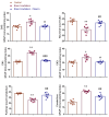
Effects of brain irradiation and niacin treatment on MDA and TNF-α levels. Data are presented as mean ± SEM. * p < 0.001 compared to the control group; # p < 0.001 compared to the brain irradiation group.
Figure 2
Figure 2
Effects of brain irradiation and niacin treatment on neurotrophic and neuroplasticity-related markers (BDNF, CREB, SIRT-1, and SIRT-6) in rat brain tissue. Data are expressed as mean ± SEM (n = 7 per group). * p < 0.001 vs. control group; # p < 0.01, ## p < 0.001 vs. brain irradiation group.
Figure 3
Figure 3
Behavioral outcomes following brain irradiation and niacin treatment. Graphs show group comparisons for sociability (time spent with stranger rat and sociability ratio), locomotor activity (ambulations in the open field), and memory performance (step-through latency in the passive avoidance test). Data are presented as mean ± SEM.* p < 0.001 vs. control; # p < 0.05, ## p < 0.01 vs. brain irradiation group.
Figure 4
Figure 4
Histological and immunohistochemical evaluations of neuronal survival (CA1, CA3, and Purkinje cell counts) and astrocytic activation (GFAP immunostaining index in CA1, CA3, and cerebellum) across groups. Brain irradiation significantly reduced neuronal counts and increased GFAP expression, indicating neurodegeneration and reactive gliosis. Niacin treatment partially restored neuronal integrity and attenuated astrocyte activation. * p < 0.01, ** p < 0.001 distinct from control groups; # p < 0.05, ## p < 0.01, ### p < 0.001 distinct from Brain Irradiation group.
Figure 5
Figure 5
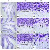
CA1 and CA3 regions of hippocampus Cresyl violet stain ×4 and ×40 magnification. (AA2): Normal Control Group female Rats CA1 and CA3 have normal pyramidal neuron (asterisk); (BB2): Brain Irradiation and saline group female rats have decreased Normal pyramidal neuron count (asterisk) and increased dysmorphological changes pyramidal neuron (arrow); (CC2): Brain Irradiation and Niacin group female rats have have increased Normal pyramidal Neuron count (asterisk), improved pyramidal neuron morphology changes (asterisk) and decreased dysmorphological changes pyramidal Neuron (arrow). (Scale bars for 1 cm = 50 μm).
Figure 6
Figure 6
CA1 and CA3 of hippocampus ×40 magnification. Astrogliosis was characterized by GFAP immunostaining (Brown staining). (AA2) Normal Control Group female Rats CA1 and CA3, (BB2) Brain Irradiation and saline group female rats have increased glial activity CA1 and CA3. (CC2) Brain Irradiation and Niacin group female rats have decreased glial activity in CA1 and CA3 (Scale bars for 1 cm = 50 μm).
Figure 7
Figure 7
Cerebellum Cresyl violet stain ×40 and ×100 magnification. (AA2), Normal Control Group female Rats cerebellum have normal Purkinje Neuron (asterisk); (BB2): Brain Irradiation and saline group female rats have decreased Normal Purkinje Neuron count (asterisk) and increased dysmorphological changes Purkinje Neuron (arrow); (CC2): Brain Irradiation and Niacin group female rats have increased Normal Purkinje Neuron count (asterisk), improved Purkinje neuron morphology changes (asterisk) and decreased dysmorphological changes Purkinje Neuron (arrow). (Scale bars for 1 cm = 50 μm).
Figure 8
Figure 8
Female rats’ cerebellum ×40 and ×100 magnification. GFAP staining (brown) revealed astrogliosis. (AA2) Control rats; (BB2) Brain Irradiation rats have enhanced glial level; (CC2) Brain Irradiation +niacin rats have reduced glial activity. Scale bars: 50 μm for ×40 magnification images (A1,A2,B1,B2,C1,C2).
Figure 9
Figure 9
Experimental procedure and timeline.

Similar articles

References

    1. Gzell C., Back M., Wheeler H., Bailey D., Foote M. Radiotherapy in Glioblastoma: The Past, the Present and the Future. Clin. Oncol. 2017;29:15–25. doi: 10.1016/j.clon.2016.09.015. - DOI - PubMed
    1. Greene-Schloesser D., Robbins M.E., Peiffer A.M., Shaw E.G., Wheeler K.T., Chan M.D. Radiation-induced brain injury: A review. Front. Oncol. 2012;2:73. doi: 10.3389/fonc.2012.00073. - DOI - PMC - PubMed
    1. Lee H.J., Kim J.S., Moon C., Son Y. Profiling of gene expression in the brain associated with anxiety-related behaviors in the chronic phase following cranial irradiation. Sci. Rep. 2022;12:13162. doi: 10.1038/s41598-022-17310-z. - DOI - PMC - PubMed
    1. Crossen J.R., Garwood D., Glatstein E., Neuwelt E.A. Neurobehavioral sequelae of cranial irradiation in adults: A review of radiation-induced encephalopathy. J. Clin. Oncol. 1994;12:627–642. doi: 10.1200/JCO.1994.12.3.627. - DOI - PubMed
    1. Meyers C.A., Brown P.D. Role and relevance of neurocognitive assessment in clinical trials of patients with CNS tumors. J. Clin. Oncol. 2006;24:1305–1309. doi: 10.1200/JCO.2005.04.6086. - DOI - PubMed

MeSH terms

LinkOut - more resources