. 2025 Jun 3;26(11):5357.
doi: 10.3390/ijms26115357.

Photodynamic Evaluation of Synthesized Chlorin-Desthiobiotin Conjugate with Chemotherapeutic Drugs in Triple-Negative Breast Cancer Cells In Vitro and in Hydra Organisms In Vivo

Affiliations

Photodynamic Evaluation of Synthesized Chlorin-Desthiobiotin Conjugate with Chemotherapeutic Drugs in Triple-Negative Breast Cancer Cells In Vitro and in Hydra Organisms In Vivo

Bailey N Rutkowski et al. Int J Mol Sci. .

Abstract

In this article, the synthesis and characterization of chlorin-based photosensitizers for potential applications in photodynamic therapy (PDT) of triple-negative breast cancer (TNBC) are described. The photodynamic efficacy of the synthesized chlorin-desthiobiotin (CDBTN) conjugate and its zinc and indium complexes were compared with the starting unconjugated precursor methyl pheophorbide, and assessed in a TNBC cell line in vitro. The chlorin-desthiobiotin complex aims to target the vitamin receptors upregulated in malignant cancer cells. The synthesized CDBTN was combined with chemotherapeutic agents (paclitaxel, cisplatin or fluorouracil) to evaluate their binary photodynamic efficacy. Cell survival assay in vitro indicated that the chlorin-vitamin conjugate CDBTN-alone and in combination with paclitaxel or fluorouracil-is photoactive against the TNBC cell line, but not when combined with cisplatin. The combination index (CI) calculated using the Chou-Talalay method indicated synergism of CDBTN and fluorouracil combination, aligning with the in vitro assay. The photodynamic cytotoxicity of CDBTN was also evaluated in vivo using the hydra as a novel model organism. This study is the first to show the use of the aquatic hydra organism in assessing photodynamic activity of the photosensitizer alone or in combination with chemotherapeutic agents. In vivo results with hydras indicated that the CDBTN-cisplatin combination is more phototoxic than CDBTN-paclitaxel or CDBTN-fluorouracil binary treatment. With the proper adjustment of concentration and light dosage, the synthesized photosensitizer can provide promising application in binary chemotherapy PDT treatment of TNBC.

Keywords: PDT; TNBC; chemophotodynamic therapy; chemotherapy; chlorin-vitamin conjugates; cisplatin; fluorouracil; hydra model organism; paclitaxel; photodynamic therapy; photosensitizers; triple-negative breast cancer.


Conflict of interest statement

The authors declare no conflict of interest.

Figures

Scheme 1
Scheme 1
Synthesis of desthiobiotin-linked chlorin derivative and its corresponding zinc and indium complexes. Reaction conditions: (i) H2N(CH2)6NHBoc, CH2Cl2; (ii) trifluoroacetic acid (TFA), CH2Cl2; (iii) desthiobiotin, DMTMM; (iv) Zn(OAc)2 in MeOH; and, (v) InCl3 in NaOAc/HOAc.
Figure 1
Figure 1
UV-vis absorption spectra in dichloromethane of chlorin-desthiobiotin conjugate (CDBTN) 3, ZnCDBTN 4, InCDBTN-Cl 5.
Figure 2
Figure 2
Molecular structures of chemotherapeutic agents.
Figure 3
Figure 3
Bar plots of cell survival assay of triple-negative BT-549 breast cancer cell line after 24 h PS (MePheo, CDBTN, InCDBTN-Cl, and ZnCDBTN) treatment: (A) in the dark, and (B) after light exposure. Cells were treated with varying concentrations (10, 25, 50, 75, and 100 nM) of PSs. Light dose = 0.96 J cm−2. Data reported are the mean ± SD of triplicate measurements in three separate experiments.
Figure 3
Figure 3
Bar plots of cell survival assay of triple-negative BT-549 breast cancer cell line after 24 h PS (MePheo, CDBTN, InCDBTN-Cl, and ZnCDBTN) treatment: (A) in the dark, and (B) after light exposure. Cells were treated with varying concentrations (10, 25, 50, 75, and 100 nM) of PSs. Light dose = 0.96 J cm−2. Data reported are the mean ± SD of triplicate measurements in three separate experiments.
Figure 4
Figure 4
Bar plots of cell survival assay of triple-negative BT-549 breast cancer cell line after 24 h PS (CDBTN, InCDBTN-Cl, and ZnCDBTN) co-treated with PTX (A) in the dark, and (B) after light exposure. Cells were treated with varying concentrations (10, 25, 50, 75, and 100 nM) of PSs. PTX concentration is 50 nM. Light dose = 0.96 J cm−2. Data reported are the mean ± SD of triplicate measurements in three separate experiments (p < 0.05, two-tailed t-test).
Figure 5
Figure 5
Bar plots of cell survival assay of triple-negative BT-549 breast cancer cell line after 24 h PS (CDBTN, InCDBTN-Cl, and ZnCDBTN) co-treated with CisPt (A) in the dark, and (B) after light exposure. Cells were treated with varying concentrations (10, 25, 50, 75, and 100 nM) of PSs. CisPt concentration was 25 μM. Light dose = 0.96 J cm−2. Data reported are the mean ± SD of triplicate measurements in three separate experiments. CDBTN at 50–75 nM showed a statistically insignificant effect upon combination with CisPt (p > 0.05, two-tailed t-test).
Figure 6
Figure 6
Bar plots of cell survival assay of triple-negative BT-549 breast cancer cell line after 24 h PS (CDBTN, InCDBTN-Cl, and ZnCDBTN) co-treated with FU (A) in the dark, and (B) after light exposure. Cells were treated with varying concentrations (10, 25, 50, 75, and 100 nM) of PSs. FU concentration was 25 μM. Light dose = 0.96 J cm−2. Data reported are the mean ± SD of triplicate measurements in three separate experiments (p < 0.05, two-tailed t-test).
Figure 7
Figure 7
Fluorescence microscopy images of fixed human triple-negative breast cancer (TNBC) BT-549 cells. Grayscale and blue fluorescence images of cells stained with Hoechst 33258. Morphology of (A) untreated unirradiated cells (sham control); then, of (B) cells alone after treatment with 2 min light, and of cells after 24 h treatment with (C) DBTN, 500 nM; (D) CDBTN (50 nM), dark; (E) MePheo, followed by 1 min light irradiation; (F) PTX, 50 nM (dark); (G) CDBTN (50 nM) + PTX (50 nM) in the dark; (H) CDBTN (50 nM) + PTX (50 nM), 30 s light exposure; (I) FU, 25 μM (dark); and PSs at 50 nM combined with FU (25 μM), followed by 1 min light exposure: (J) CDBTN; (K) ZnCDBTN; and (L) InCDBTN-Cl, respectively. Apoptosis characterized by chromatin condensation and cell shrinkage is evident upon treatment with PTX (F) and with CDBTN with FU (J). Light exposure for 30 s and 1 min corresponds to light doses of 0.48 and 0.96 J cm−2, respectively. Scale bars are the same for all images as in the first figure. Objective used was 20X.
Figure 8
Figure 8
Ultrastructure TEM images of human triple-negative breast cancer (TNBC) BT-549 cells: (A) untreated irradiated for 1 min; (B) CDBTN-treated and unirradiated; (C) CDBTN-treated with 30 s light treatment; (D) PTX-treated treatment, with 30 s light treatment; (E) CDBTN-treated and co-treated with PTX without and (F) with 30 s light treatment; (G) with FU without, and (H) with 30 s light treatment; and, (I) with CisPt without and (J) 30 s light treatment. Irregularly shaped nuclear membrane is apparent in treated cells in the presence of light. Light dose in 30 s = 0.48 J cm−2, and in 1 min = 0.96 J cm−2. Concentrations: PS = 50 nM.
Figure 9
Figure 9
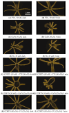
Morphological changes of hydra organisms treated with synthesized PSs: (A) Green hydra in the dark; (B) hydra being fed with brine shrimps; (C) hydra, irradiated for 2 min; (DF) CDBTN, dark and irradiated; (GI) InCDBTN-CL, dark and irradiated; (JL) ZnCDBTN, dark and irradiated. Light dose in 2 min = 1.92 J cm−2 and 5 min = 4.8 J cm−2. Concentrations: PS = 1.0 μM. Scale bars are the same for all images as in the first figure. Objective used was 10X.
Figure 10
Figure 10
Morphological changes of hydra organisms treated with chemotherapeutic drugs and PS: (A) PTX in the dark; (B) PTX, irradiated; (C) CisPt in the dark; (D) CisPt, irradiated; (E) 5-FU in the dark; (F) FU, irradiated; (G) co-treatment with CDBTN and PTX in the dark; (H) co-treatment with CDBTN and PTX, irradiated; (I) co-treatment with CDBTN and CisPt in the dark; (J) co-treatment with CDBTN and CisPt, irradiated; (K) co-treatment with CDBTN and FU in the dark; and (L) co-treatment with CDBTN and PTX, irradiated. An irregularly shaped nuclear membrane is apparent in treated cells in the presence of light. Light dose in 5 min = 4.8 J cm−2. Concentrations: PTX = 50 nM; CisPt = 25 μM; FU = 25 μM; CDBTN = 50 nM. Scale bars are the same for all images as in the first figure. Objective used was 10X.

Similar articles

References

    1. Wang X., Peng J., Meng C., Feng F. Recent Advances for enhanced photodynamic therapy: From new mechanisms to innovative strategies. Chem. Sci. 2024;15:12234–12257. doi: 10.1039/D3SC07006A. - DOI - PMC - PubMed
    1. Obaid G., Celli J.P., Broekgaarden M., Bulin A.-L., Uusimaa P., Pogue B., Hasan T., Huang H.-C. Engineering photodynamics for treatment, priming and imaging. Nat. Rev. Bioeng. 2024;2:752–769. doi: 10.1038/s44222-024-00196-z. - DOI - PMC - PubMed
    1. Allamyradov Y., ben Yosef J., Annamuradov B., Ateyeh M., Street C., Whipple H., Er A.O. Photodynamic Therapy Review: Past, Present, Future, Opportunities and Challenges. Photochem. 2024;4:434–461. doi: 10.3390/photochem4040027. - DOI
    1. Jiang W., Liang M., Lei Q., Li G., Wu S. The Current Status of Photodynamic Therapy in Cancer Treatment. Cancers. 2023;15:585. doi: 10.3390/cancers15030585. - DOI - PMC - PubMed
    1. Ji B., Wei M., Yang B. Recent advances in nanomedicines for photodynamic therapy (PDT)-driven cancer immunotherapy. Theranostics. 2022;12:434–458. doi: 10.7150/thno.67300. - DOI - PMC - PubMed

MeSH terms

LinkOut - more resources