. 2025 Jun 3;26(11):5364.
doi: 10.3390/ijms26115364.

Colonizing Bacteria Aggravate Inflammation, Cytotoxicity and Immune Defense During Influenza A Virus Infection

Affiliations

Colonizing Bacteria Aggravate Inflammation, Cytotoxicity and Immune Defense During Influenza A Virus Infection

Liane Giebeler et al. Int J Mol Sci. .

Abstract

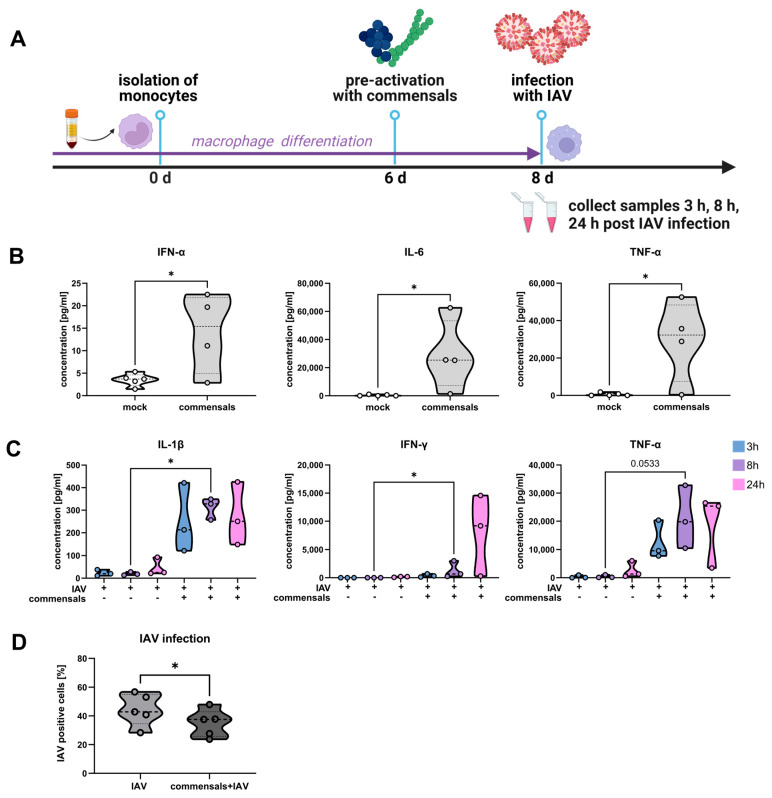
A diverse bacterial community colonizes the respiratory system, including commensals such as Staphylococcus epidermidis (S. epidermidis) and Streptococcus salivarius (S. salivarius), as well as facultative pathogens like Staphylococcus aureus (S. aureus). This study aimed to establish a colonized cell culture model to investigate the impact of these bacteria on influenza A virus (IAV) infection. Respiratory epithelial cells were exposed to S. epidermidis, S. salivarius, or S. aureus, using either live or heat-inactivated bacteria, followed by IAV infection. Cell integrity was assessed microscopically, cytotoxicity was measured via LDH assay, and inflammatory responses were analyzed through cytokine expression. Additionally, macrophage function was examined in response to bacterial colonization and IAV infection. While commensals maintained epithelial integrity for 48 h, S. aureus induced severe cell damage and death. The most pronounced epithelial destruction was caused by coinfection with S. aureus and IAV. Notably, commensals did not confer protection against IAV but instead enhanced epithelial inflammation. These effects were dependent on live bacteria, as inactivated bacteria had no impact. However, prior exposure to S. epidermidis and S. salivarius improved macrophage-mediated immune responses against IAV. These findings suggest that while individual commensals do not directly protect epithelial cells, they may contribute to immune training and enhance lung defense mechanisms.

Keywords: cell culture model; commensal bacteria; cytotoxicity; facultative pathogenic bacteria; influenza A virus; macrophages; respiratory microbiome.


Conflict of interest statement

The authors declare no conflicts of interest.

Figures

Figure 1
Figure 1
S. aureus harms the integrity of the epithelial cell layer compared to S. epidermidis and S. salivarius. (A) Study design of the colonization of Calu-3 cells with live or inactivated commensal (S. salivarius and S. epidermidis) and opportunistic bacteria (S. aureus). Light microscopy pictures of Calu-3 cells inoculated with either (B) S. salivarius and S. epidermidis (0.00001 MOI for live commensals; 1.85 × 10⁶ bacteria/well for inactivated S. salivarius and 2.1 × 10⁶ bacteria/well for inactivated S. epidermidis) or (C) S. aureus (0.00002 MOI for live S. aureus; 2 × 10⁸ bacteria/well for inactivated S. aureus). The cells were washed twice with PBS at 4 and 8 h post-inoculation (p. io.), respectively, to reduce the amount of non-internalized bacteria. Microscopic analysis was performed 32 or 48 h p. io. as indicated. Scale bars represent 200 µm.
Figure 2
Figure 2
Colonization of the epithelial layer with live bacteria does not protect from an influenza virus infection, but adds to the epithelial damage. (A) Study design of the colonization of Calu-3 cells with live or inactivated commensal and opportunistic bacteria, followed by an influenza A virus infection (IAV). (B) Light microscopy pictures of Calu-3 cells inoculated with either S. salivarius and S. epidermidis (0.00001 MOI for live commensals; 1.85 × 10⁶ bacteria/well for inactivated S. salivarius and 2.1 × 10⁶ bacteria/well for inactivated S. epidermidis) or (C) S. aureus (0.00002 MOI for live S. aureus; 2 × 10⁸ bacteria/well for inactivated S. aureus). The cells were washed twice with PBS at 4 and 8 h p. io., respectively, to reduce the amount of non-internalized bacteria. Infection with IAV was performed 24 h p. io. Microscopic analysis was performed 32 h or 48 h p. io. Scale bars represent 200 µm.
Figure 3
Figure 3
Live S. aureus (but not commensal bacteria) increases the LDH release of epithelial cells. Calu-3 cells were inoculated with either (A) S. salivarius and S. epidermidis or (B) S. aureus as live or inactivated bacteria as indicated, washed twice with PBS at 4 and 8 h, and infected with IAV (1 MOI) at 24 h p. io. Samples were taken at 48 h p. io. Cell death rate was assessed with LDH assays 48 h p.io. Data shown represent the means ± SD of at least three independent experiments with two technical replicates. Statistical analysis was performed with Kruskal–Wallis test and Dunn’s multiple comparison test. * p < 0.05, ** p < 0.01, *** p < 0.001.
Figure 4
Figure 4
Live commensal bacteria induce the expression of interleukin-6 (IL-6) and -8 (IL-8) in epithelial cells. Calu-3 cells were inoculated with either S. salivarius and S. epidermidis or S. aureus as live or inactivated bacteria, as indicated, washed twice with PBS at 4 and 8 h, and infected with IAV (1 MOI) at 24 h p. io. RNA was extracted at 32 h p. io. with either live (left panel) or inactivated (right panel) commensal bacteria, followed by cDNA synthesis and qRT-PCR for IL-6 (A) and IL-8 (B). Data represent the median of at least three independent experiments with two technical replicates. Statistical significance was determined by Kruskal–Wallis test and Dunn’s multiple comparison test. * p < 0.05; ** p < 0.01; **** p < 0.0001.
Figure 5
Figure 5
Commensal bacteria enhance the viral titers in the supernatants of epithelial cells. Calu-3 cells were inoculated with control medium compared to commensals (S. salivarius and S. epidermidis) as live or inactivated bacteria, as indicated, washed twice with PBS at 4 and 8 h, and infected with IAV (1 MOI) at 24 h p. io. IAV titers were determined by standard plaque assays at 32 h p. io. for live (left panel) or inactivated (right panel) commensals. Titers are shown in percent with the mean of the single infection set as 100%. Data represent means + SD of at least three independent experiments with two technical replicates. Statistical significance was determined by Mann–Whitney test ** p < 0.01.
Figure 6
Figure 6
Commensal bacteria pre-activate macrophages and enhance the clearance of IAV infection. (A) Study design of monocyte-derived macrophages that were pre-stimulated with the inactivated commensal bacteria S. epidermidis (MOI 0.5) and S. salivarius (MOI 0.5) for 2 d prior to the infection with IAV (MOI 1). (B) The antiviral cytokine response was measured in the supernatants of monocyte-derived macrophages with or without pre-stimulation with inactivated commensals for 2 d and (C) with subsequent IAV infection for 3 h, 8 h, and 24 h via multiplexed immunoassay (n = 3; Kruskal–Wallis test and Dunn’s multiple comparison test). (D) IAV-positive macrophages were quantified 24 h post IAV infection by flow cytometry with or without pre-stimulation with inactivated commensal bacteria (n = 5, paired t-test). * p < 0.05.

References

    1. Wypych T.P., Wickramasinghe L.C., Marsland B.J. The influence of the microbiome on respiratory health. Nat. Immunol. 2019;20:1279–1290. doi: 10.1038/s41590-019-0451-9. - DOI - PubMed
    1. Otto M. Staphylococcus epidermidis—the ‘accidental’ pathogen. Nat. Rev. Microbiol. 2009;7:555–567. doi: 10.1038/nrmicro2182. - DOI - PMC - PubMed
    1. Delorme C., Abraham A.L., Renault P., Guédon E. Genomics of Streptococcus salivarius, a major human commensal. Infect. Genet. Evol. 2015;33:381–392. doi: 10.1016/j.meegid.2014.10.001. - DOI - PubMed
    1. Oberbach A., Schlichting N., Hagl C., Lehmann S., Kullnick Y., Friedrich M., Köhl U., Horn F., Kumbhari V., Löffler B., et al. Four decades of experience of prosthetic valve endocarditis reflect a high variety of diverse pathogens. Cardiovasc. Res. 2023;119:410–428. doi: 10.1093/cvr/cvac055. - DOI - PubMed
    1. Liu Q., Liu Q., Meng H., Lv H., Liu Y., Liu J., Wang H., He L., Qin J., Wang Y., et al. Staphylococcus epidermidis Contributes to Healthy Maturation of the Nasal Microbiome by Stimulating Antimicrobial Peptide Production. Cell Host Microbe. 2020;27:68–78.e5. doi: 10.1016/j.chom.2019.11.003. - DOI - PMC - PubMed

MeSH terms

LinkOut - more resources