

Tacrolimus Modulates TGF-β Signaling-Related Genes and MicroRNAs in Human Retinal Pigment Epithelial Cells Activated by Lipopolysaccharide
- PMID: 40508210
- PMCID: PMC12156939
- DOI: 10.3390/ijms26115402
Tacrolimus Modulates TGF-β Signaling-Related Genes and MicroRNAs in Human Retinal Pigment Epithelial Cells Activated by Lipopolysaccharide
Abstract
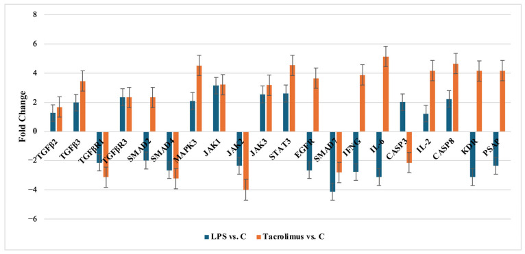
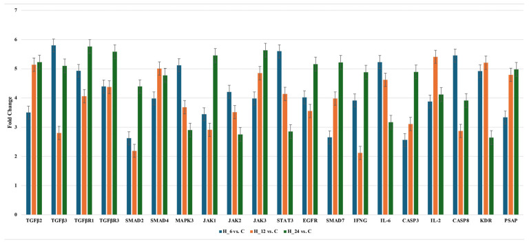
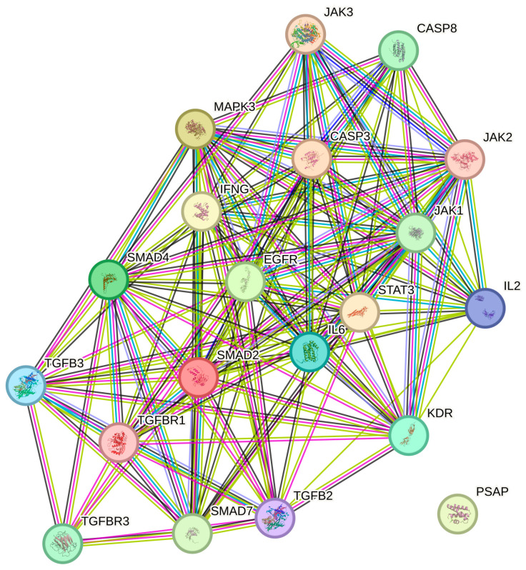
The retinal pigment epithelium (RPE) plays a crucial role in maintaining retinal homeostasis, and dysregulation of the transforming growth factor-beta (TGF-β) signaling pathways contributes to retinal fibrosis and inflammatory diseases, including proliferative vitreoretinopathy (PVR). Tacrolimus (FK506), an immunosuppressant, has shown potential antifibrotic properties, but its effects on TGF-β-related genes and microRNAs (miRNAs) in RPE cells remain unclear. Human RPE (H-RPE) cells were treated with lipopolysaccharide (LPS) to induce inflammation and subsequently exposed to tacrolimus. Gene and miRNA expression profiling related to TGF-β signaling pathways were conducted using microarrays, followed by Quantitative Reverse-Transcription Polymerase Chain Reaction (RT-qPCR) validation. Protein levels were assessed via enzyme-linked immunosorbent assay (ELISA), and interactions were analyzed using STRING database network analysis. Tacrolimus modulated key components of the TGF-β pathway, upregulating TGF-β2, TGF-β3, SMAD2, and SMAD4 while downregulating TGF-βR1 and SMAD7. JAK/STAT and MAPK pathways were also affected, indicating broad regulatory effects. miRNA profiling identified hsa-miR-200a-3p, hsa-miR-589-3p, hsa-miR-21, and hsa-miR-27a-5p as key regulators. STRING analysis confirmed strong functional interactions within the TGF-β network. In conclusion, tacrolimus modulates both canonical (upregulation of SMAD2/4 and downregulation of SMAD7) and non-canonical (JAK/STAT and MAPK) TGF-β signaling pathways in LPS-stimulated RPE cells. These changes collectively suggest a dual anti-inflammatory and anti-fibrotic effect. The increased TGF-β2 and decreased SMAD7 levels, alongside altered miRNA expression (e.g., downregulation of miR-200a-3p), indicate that tacrolimus may inhibit key profibrotic mechanisms underlying PVR. These findings support the potential therapeutic repurposing of tacrolimus in PVR and warrant further in vivo validation.
Keywords: lipopolysaccharide (LPS); proliferative vitreoretinopathy; retinal pigment epithelium; tacrolimus; transforming growth factor-beta (TGF-β) signaling pathway.
Conflict of interest statement
The authors declare no conflicts of interest.
Figures
Similar articles
-
Evaluation of Gene Expression and the Regulatory Role of microRNAs Related to the Mitogen-Activated Protein Kinase Signaling Pathway in Human Retinal Pigment Epithelial Cells Treated With Lipopolysaccharide A and Tacrolimus.Mediators Inflamm. 2025 Jul 1;2025:8586711. doi: 10.1155/mi/8586711. eCollection 2025. Mediators Inflamm. 2025. PMID: 40630080 Free PMC article.
-
Transforming growth factor β-related genes in human retinal pigment epithelial cells after tacrolimus treatment.Pharmacol Rep. 2016 Oct;68(5):969-74. doi: 10.1016/j.pharep.2016.04.020. Epub 2016 May 11. Pharmacol Rep. 2016. PMID: 27372923
-
Blockade of Jagged/Notch pathway abrogates transforming growth factor β2-induced epithelial-mesenchymal transition in human retinal pigment epithelium cells.Curr Mol Med. 2014 May;14(4):523-34. doi: 10.2174/1566524014666140331230411. Curr Mol Med. 2014. PMID: 24694299 Review.
-
Pirfenidone inhibits transforming growth factor-β1-induced fibrogenesis by blocking nuclear translocation of Smads in human retinal pigment epithelial cell line ARPE-19.Mol Vis. 2012;18:1010-20. Epub 2012 Apr 21. Mol Vis. 2012. PMID: 22550395 Free PMC article.
-
Differential regulation of microRNA-146a and microRNA-146b-5p in human retinal pigment epithelial cells by interleukin-1β, tumor necrosis factor-α, and interferon-γ.Mol Vis. 2013 Apr 3;19:737-50. Print 2013. Mol Vis. 2013. PMID: 23592910 Free PMC article.
References
MeSH terms
Substances
LinkOut - more resources
Full Text Sources
Research Materials
Miscellaneous
