Review
. 2025 May 26;17(11):1475.
doi: 10.3390/polym17111475.

Regenerative Endodontic Therapies: Harnessing Stem Cells, Scaffolds, and Growth Factors

Affiliations
Review

Regenerative Endodontic Therapies: Harnessing Stem Cells, Scaffolds, and Growth Factors

Rosana Farjaminejad et al. Polymers (Basel). .

Abstract

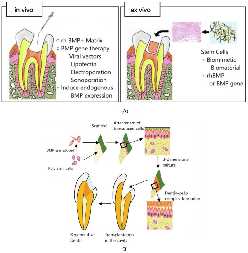
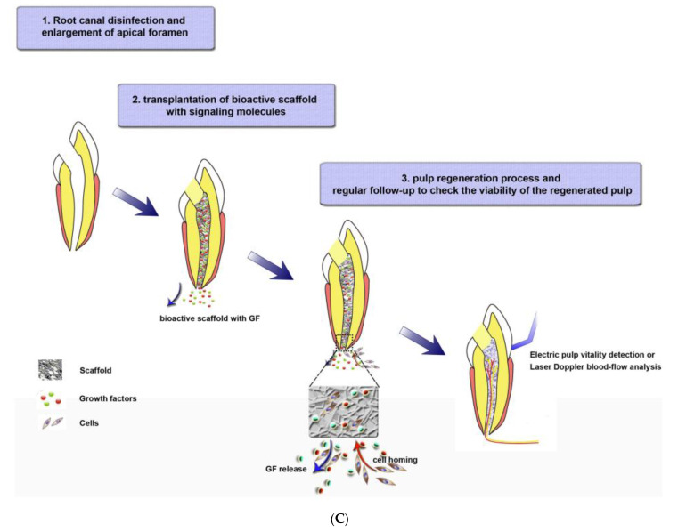
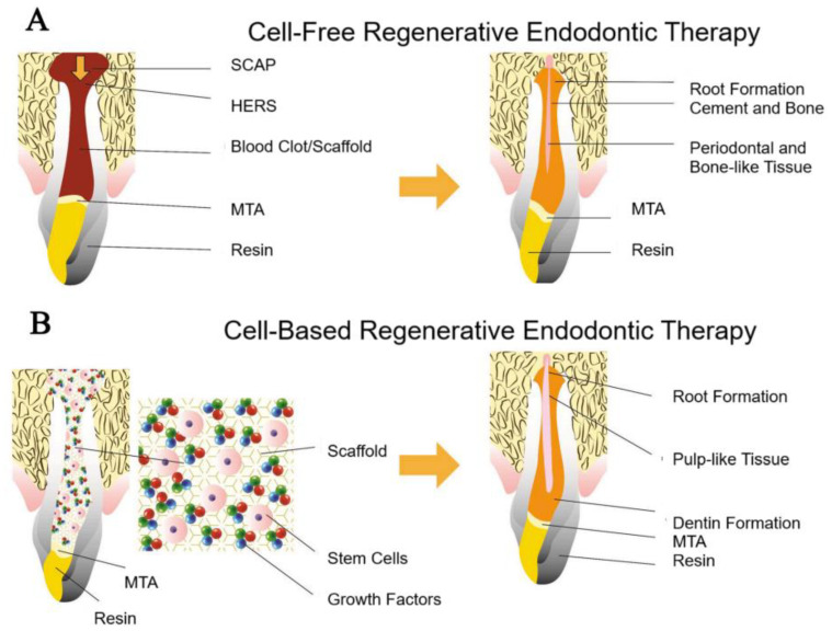
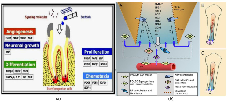
Regenerative Endodontic Therapies (RETs) offer transformative potential by leveraging polymer-based scaffolds, stem cells, and growth factors to regenerate damaged dental pulp tissue, thereby restoring tooth vitality and prolonging tooth function. While conventional treatments focus on infection control, they often compromise the structural and biological integrity of the tooth. RETs, in contrast, aim to restore the natural function of the pulp-dentin complex by promoting cellular regeneration and immune modulation. In this context, biodegradable polymers-such as collagen, gelatin methacryloyl (GelMA), and synthetic alternatives-serve as scaffolding materials that mimic the extracellular matrix, support cell attachment and proliferation, and enable localized delivery of bioactive factors. Together, the tissue engineering triad-polymer-based scaffolds, stem cells, and signaling molecules-facilitates root development, apical closure, and increased fracture resistance. Recent innovations in polymeric scaffold design, including injectable hydrogels and 3D bioprinting technologies, have enhanced clinical translation by enabling minimally invasive and patient-specific RETs. Despite progress, challenges such as immune compatibility, scaffold degradation rates, and the standardization of clinical protocols remain. RETs, thus, represent a paradigm shift in dental care, aligning with the body's intrinsic healing capacity and offering improved long-term outcomes for patients.

Keywords: 3D bioprinting; collagen; dental; dental pulp stem cells; regeneration; transforming growth factor beta.


Conflict of interest statement

The authors declare no conflicts of interest.

Figures

Figure 1
Figure 1
(A) Three therapeutic strategies have been proposed for treating endodontic and periodontal diseases using dental mesenchymal stem cells (MSCs): (a) dental tissue regeneration through the classic tissue engineering model, which involves the use of dental MSCs combined with supporting biomaterial scaffolds and growth factors; (b) dental tissue regeneration via scaffold-free tissue engineering approaches; and (c) a cell-free strategy that promotes dental tissue regeneration using conditioned medium (CM) containing exosomes and/or extracellular vesicles (EVs) secreted by dental MSCs. (B) Pulp regeneration process using decellularized dental pulp tissue matrix. The cycle starts from the decellularization of pulp tissue to form a dental pulp tissue matrix scaffold, which is then delivered into the tooth structure for regeneration. Adopted from [10,12].
Figure 2
Figure 2
Schematic illustrating the integration of stem cells, scaffolds, and growth factors in RE for pulp–dentin regeneration. Depiction of various scaffolds used in dental TE, including natural and synthetic polymers, that support dental pulp regeneration by interacting with stem cells and growth factors.
Figure 3
Figure 3
The application of irrigates and medicaments to a dentin matrix releases bioactive molecules, enhancing chemotaxis, angiogenesis, neurogenesis, and differentiation, which together stimulate odontoblasts and support regeneration within the dentin matrix. Adopted from [47].
Figure 4
Figure 4
Injectable scaffolds show significant potential for minimally invasive pulp regeneration, promoting faster recovery and reducing complications. The scaffold is injected into the treatment site, where it promotes tissue regeneration by modulating the immune response, balancing pro- and anti-inflammatory macrophages. Evaluation methods such as micro-CT, histology, and RNA sequencing are used to assess the outcome of the treatment. Adopted from [58].
Figure 5
Figure 5
(A) Dentin regeneration techniques: in vivo methods use rhBMP, gene therapy, and electroporation to induce BMP expression; ex vivo approaches involve transplanting odontoblasts or stem cells with biomaterials and BMP genes into dentin matrices. (B) Regeneration process illustrating the steps from BMP transduction of pulp stem cells, their attachment to scaffolds, and the formation of a dentin–pulp complex in a 3-dimensional culture, ultimately leading to the regeneration and transplantation of dentin in dental cavities. (C) RE via cell homing strategy: (1) The process begins with disinfecting the root canal and enlarging the apical foramen to prepare for regeneration. (2) A bioactive scaffold is then implanted into the cleaned canal, releasing growth factors that attract cells critical for tissue formation. (3) As these cells migrate, proliferate, and differentiate within the scaffold, they contribute to the development of vital dental structures such as blood vessels, nerves, and dentin. Regular follow-ups are essential to monitor the health and integration of the regenerated pulp. Adopted from [43,47,53].
Figure 5
Figure 5
(A) Dentin regeneration techniques: in vivo methods use rhBMP, gene therapy, and electroporation to induce BMP expression; ex vivo approaches involve transplanting odontoblasts or stem cells with biomaterials and BMP genes into dentin matrices. (B) Regeneration process illustrating the steps from BMP transduction of pulp stem cells, their attachment to scaffolds, and the formation of a dentin–pulp complex in a 3-dimensional culture, ultimately leading to the regeneration and transplantation of dentin in dental cavities. (C) RE via cell homing strategy: (1) The process begins with disinfecting the root canal and enlarging the apical foramen to prepare for regeneration. (2) A bioactive scaffold is then implanted into the cleaned canal, releasing growth factors that attract cells critical for tissue formation. (3) As these cells migrate, proliferate, and differentiate within the scaffold, they contribute to the development of vital dental structures such as blood vessels, nerves, and dentin. Regular follow-ups are essential to monitor the health and integration of the regenerated pulp. Adopted from [43,47,53].
Figure 6
Figure 6
Schematic diagram illustrating the two Regenerative Endodontic Therapy approaches with the use of biomaterial scaffolds. (A) Cell-free Regenerative Endodontic Therapy: a blood clot or scaffold is introduced into the canal space, leading to the regeneration of periodontal and bone-like tissue. (B) Cell-based Regenerative Endodontic Therapy: stem cells, scaffolds, and growth factors are placed in the canal space, resulting in the regeneration of pulp-like tissue. The key difference is that the cell-based approach promotes true pulp regeneration, whereas the cell-free approach encourages repair tissue formation. SCAP: stem cells from the apical papilla; HERS: Hertwig’s epithelial root sheath; MTA: mineral trioxide aggregate. Adapted with permission [69].
Figure 7
Figure 7
(a) Depicts the interaction of growth factors with stem cells and pericytes in dental pulp regeneration, emphasizing angiogenesis and tissue formation (b) Showcases a detailed schematic of regenerative endodontic therapy, with a focus on growth factor deployment, stem cell interaction, and scaffold implantation to foster dental pulp regeneration [174,175].

Similar articles

References

    1. Soares D.G., Bordini E.A., Swanson W.B., de Souza Costa C.A., Bottino M.C. Platform technologies for regenerative endodontics from multifunctional biomaterials to tooth-on-a-chip strategies. Clin. Oral Investig. 2021;25:4749–4779. doi: 10.1007/s00784-021-04013-4. - DOI - PMC - PubMed
    1. Boyd R.C. Wiggs’s Veterinary Dentistry: Principles and Practice. John Wiley & Sons; Hoboken, NJ, USA: 2019. Basic endodontic therapy; pp. 311–334.
    1. Howard D., Buttery L.D., Shakesheff K.M., Roberts S.J. Tissue engineering: Strategies, stem cells and scaffolds. J. Anat. 2008;213:66–72. doi: 10.1111/j.1469-7580.2008.00878.x. - DOI - PMC - PubMed
    1. Widbiller M., Schmalz G. Endodontic regeneration: Hard shell, soft core. Odontology. 2021;109:303–312. doi: 10.1007/s10266-020-00573-1. - DOI - PMC - PubMed
    1. Al-Helou N. In: Contemporary Endodontics for Children and Adolescents. Fuks A.B., Moskovitz M., Tickotsky N., editors. Springer; Berlin, Germany: 2023.

LinkOut - more resources