

Optimization of Bond Strength Between Heat-Polymerized PMMA and Contemporary CAD/CAM Framework Materials: A Comparative In Vitro Study
- PMID: 40508730
- PMCID: PMC12157249
- DOI: 10.3390/polym17111488
Optimization of Bond Strength Between Heat-Polymerized PMMA and Contemporary CAD/CAM Framework Materials: A Comparative In Vitro Study
Abstract
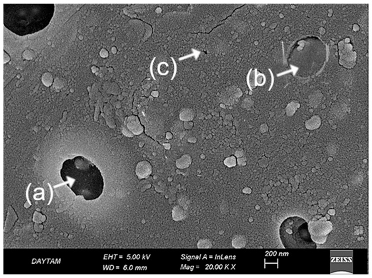
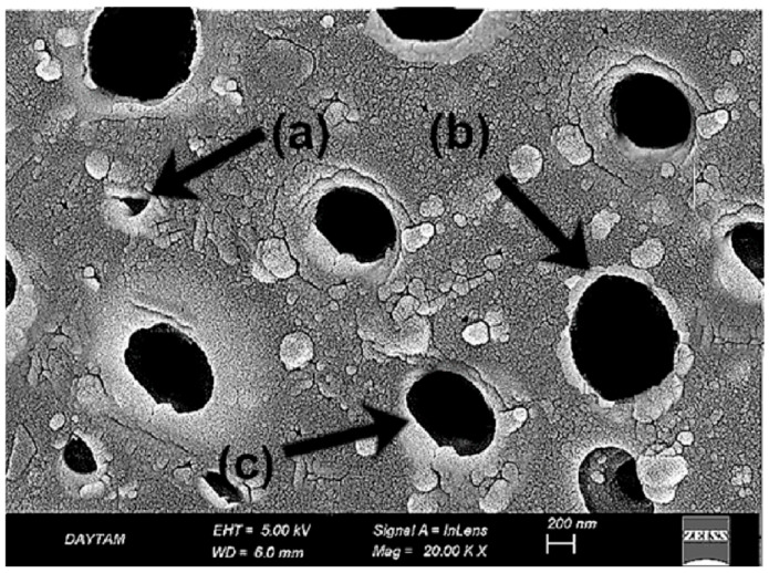
This study aimed to comparatively evaluate the effects of various surface treatment protocols on the shear bond strength (SBS) between heat-polymerized polymethyl methacrylate (PMMA) and different CAD/CAM framework materials, including cobalt-chromium (Co-Cr) alloys, ceramic particle-reinforced polyetheretherketone (PEEK), and glass fiber-reinforced composite resin (FRC). A total of 135 disc-shaped specimens were prepared from Co-Cr, PEEK, and FRC materials. Surface treatments specific to each material, including airborne-particle abrasion, sulfuric acid etching, laser irradiation, plasma activation, and primer application, were applied. PMMA cylinders were polymerized onto the treated surfaces, and all specimens were subjected to 30,000 thermal cycles. SBS values were measured using a universal testing machine, and the failure modes were classified. The normality of data distribution was assessed using the Kolmogorov-Smirnov test, and the homogeneity of variances was evaluated using Levene's test. Group comparisons were performed using the Kruskal-Wallis test, and Dunn's post hoc test with Bonferroni correction was applied in cases where significant differences were detected (α = 0.05). The highest SBS values (~27-28 MPa) were obtained in the Co-Cr group and in the PEEK groups treated with sulfuric acid and primer. In contrast, the PEEK group with additional laser treatment exhibited a lower SBS value. The untreated PEEK group showed significantly lower SBS (~3.9 MPa) compared to all other groups. The Trinia groups demonstrated intermediate SBS values (16.5-17.4 MPa), which exceeded the clinically acceptable threshold of 10 MPa. SEM observations revealed material- and protocol-specific surface responses; plasma-treated specimens maintained topographic integrity, whereas laser-induced surfaces showed localized degradation, particularly following dual-step protocols. Fracture mode analysis indicated that higher SBS values were associated with cohesive or mixed failures. SEM observations suggested that plasma treatment preserved surface morphology more effectively than laser treatment. This study highlights the importance of selecting material-specific surface treatments to optimize bonding between CAD/CAM frameworks and PMMA. Sulfuric acid and primer provided strong adhesion for PEEK, while the addition of laser or plasma offered no further benefit, making such steps potentially unnecessary. Trinia frameworks also showed acceptable performance with conventional treatments. These findings reinforce that simplified conditioning protocols may be clinically sufficient, and indicate that FRC materials like Trinia should be more fully considered for their broader clinical potential in modern CAD/CAM-based prosthetic planning.
Keywords: CAD/CAM frameworks; polymer composites; surface properties.
Conflict of interest statement
The author declares no conflicts of interest.
Figures


Similar articles
-
Bond strength comparison of chemically activated hard reline materials on CAD-CAM milled and conventional heat-polymerized PMMA denture bases.J Prosthet Dent. 2025 Mar;133(3):889.e1-889.e9. doi: 10.1016/j.prosdent.2024.12.001. Epub 2025 Jan 2. J Prosthet Dent. 2025. PMID: 39753483
-
Effect of Surface Treatments and Thermal Aging on Bond Strength Between Veneering Resin and CAD/CAM Provisional Materials.Polymers (Basel). 2025 Feb 20;17(5):563. doi: 10.3390/polym17050563. Polymers (Basel). 2025. PMID: 40076056 Free PMC article.
-
Effect of various solvents on the repairability of aged CAD/CAM provisional restorative materials with flowable resin composite: an in vitro study.BMC Oral Health. 2025 Mar 11;25(1):368. doi: 10.1186/s12903-025-05731-x. BMC Oral Health. 2025. PMID: 40069739 Free PMC article.
-
Effect of surface treatment and resin cement type on the bond strength of polyetheretherketone to lithium disilicate ceramic.BMC Oral Health. 2024 May 2;24(1):513. doi: 10.1186/s12903-024-04269-8. BMC Oral Health. 2024. PMID: 38698366 Free PMC article.
-
Surface modification of zirconia or lithium disilicate-reinforced glass ceramic by laser texturing to increase the adhesion of prosthetic surfaces to resin cements: an integrative review.Clin Oral Investig. 2023 Jul;27(7):3331-3345. doi: 10.1007/s00784-023-05016-z. Epub 2023 Apr 17. Clin Oral Investig. 2023. PMID: 37069409 Free PMC article. Review.
Cited by
-
Shear Bond Strength of Self-Adhesive and Self-Etching Resin Cements to Dentin for Indirect Restorations.J Funct Biomater. 2025 Aug 12;16(8):289. doi: 10.3390/jfb16080289. J Funct Biomater. 2025. PMID: 40863309 Free PMC article.
References
-
- Jain R., Takkar R., Jain G., Takkar R., Deora N., Jain R. CAD-CAM the future of digital dentistry: A review. Ann. Prosthodont. Restor. Dent. 2016;2:33–36.
-
- Luthardt R., Weber A., Rudolph H., Schöne C., Quaas S., Walter M. Design and production of dental prosthetic restorations: Basic research on dental CAD/CAM technology. Int. J. Comput. Dent. 2002;5:165–176. - PubMed
-
- Curinga M.R.S., Ribeiro A.K.C., de Moraes S.L.D., do Egito Vasconcelos B.C., Carreiro A.d.F.P., Pellizzer E.P. Mechanical properties and accuracy of removable partial denture frameworks fabricated by digital and conventional techniques: A systematic review. J. Prosthet. Dent. 2025;133:85–95. doi: 10.1016/j.prosdent.2023.01.032. - DOI - PubMed
LinkOut - more resources
Full Text Sources
Miscellaneous
