. 2025 May 29;17(11):1514.
doi: 10.3390/polym17111514.

Anti-Inflammatory and Osteogenic Effect of Phloroglucinol-Enriched Whey Protein Isolate Fibrillar Coating on Ti-6Al-4V Alloy

Affiliations

Anti-Inflammatory and Osteogenic Effect of Phloroglucinol-Enriched Whey Protein Isolate Fibrillar Coating on Ti-6Al-4V Alloy

Anna Mieszkowska et al. Polymers (Basel). .

Abstract

Biomaterials play a crucial role in the long-term success of bone implant treatment. The accumulation of bacterial biofilm on the implants induces inflammation, leading to implant failure. Modification of the implant surface with bioactive molecules is one of the strategies to improve biomaterial compatibility and limit inflammation. In this study, whey protein isolate (WPI) fibrillar coatings were used as a matrix to incorporate biologically active phenolic compound phloroglucinol (PG) at different concentrations (0.1% and 0.5%) on titanium alloy (Ti6Al4V) scaffolds. Successful Ti6Al4V coatings were validated by X-ray photoelectron spectroscopy (XPS), showing a decrease in %Ti and increases in %C, %N, and %O, which demonstrate the presence of the protein layer. The biological activity of PG-enriched WPI (WPI/PG) coatings was assessed using bone-forming cells, human bone marrow-derived mesenchymal stem cells (BM-MSCs). WPI/PG coatings modulated the behavior of BM-MSCs but did not have a negative impact on cell viability. A WPI with higher concentrations of PG increased gene expression relative to osteogenesis and reduced the pro-inflammatory response of BM-MSCs after biofilm stimulation. Autoclaving reduced WPI/PG bioactivity compared to filtration. By using WPI/PG coatings, this study addresses the challenge of improving osteogenic potential while limiting biofilm-induced inflammation at the Ti6Al4V surface. These coatings represent a promising strategy to enhance implant bioactivity.

Keywords: Ti6Al4V; biofilm; fibrillar coating; inflammation; osseointegration; osteogenesis; phenolic coating; stem cells; whey protein isolate.


Conflict of interest statement

The authors declare no conflicts of interest. The funders had no role in the design of this study; in the collection, analyses, or interpretation of the data; in the writing of the manuscript; or in the decision to publish the results.

Figures

Figure 1
Figure 1
Diagram of the WPI coating process with PG. Three different coating methods are illustrated: (1) WPI coating only, (2) WPI + PG coating autoclaved together, and (3) WPI + PG coating with PG added after sterilization of the WPI coating. Ti: titanium alloy Ti6Al4V; WPI: whey protein isolate; PG: phloroglucinol.
Figure 2
Figure 2
Atomic composition of uncoated Ti and WPI coatings without PG, with 0.1% of PG and 0.5% of PG. [WPI + PG 0.1%] autoclave and [WPI + PG 0.5%] autoclave mean that the coatings containing PG were autoclaved. [WPI] autoclave + PG 0.1% and [WPI] autoclave + PG 0.5% mean that only the WPI coating was autoclaved; PG was added afterwards using a sterile filter and syringe. Error bars represent SD. Ti: titanium alloy Ti6Al4V; WPI: whey protein isolate; PG: phloroglucinol.
Figure 3
Figure 3
(a) Representative scanning electron micrograph of a multispecies biofilm formed on a glass coverslip, subsequently used in the co-culture model with BM-MSCs. Arrows in the SEM image indicate examples of four different bacterial species: S. mitis (blue arrow), F. nucleatum (green arrow), P. gingivalis (yellow arrow), and A. actinomycetemcomitans (orange arrow). (b) The number of each bacterial species quantified using the qPCR method and calculated per 100 ng of DNA isolated from the biofilm. (c) The results of bacterial quantification presented as the percentage (%) of the total number of bacteria in the biofilm.
Figure 4
Figure 4
Representative SEM images of unstimulated and biofilm-stimulated BM-MSCs on (a,g) uncoated Ti6Al4V; (b,h) WPI; (c,i) [WPI + PG 0.1%] autoclave; (d,j) [WPI + PG 0.5%] autoclave; (e,k) [WPI] autoclave + PG 0.1%; and (f,l) [WPI] autoclave + PG 0.5% coatings on Ti6Al4V. Ellipses indicate unstimulated cells (orange) and biofilm-stimulated cells (green) and white arrows show particles of Ti6Al4V powder. Scale bar: 20 μm. Ti: titanium alloy Ti6Al4V; WPI: whey protein isolate; PG: phloroglucinol.
Figure 5
Figure 5
Metabolic activity of unstimulated and biofilm-stimulated BM-MSCs after 48 h using MTT test. BM-MSCs were stimulated with biofilm for 2 h, and metabolic activity was analyzed directly after biofilm stimulation. The results are shown as mean (n = 4, two technical repetitions), and bars represent SEM. Significant differences for unstimulated vs. biofilm-stimulated BM-MSCs are indicated with & (p < 0.05) and && (p < 0.01). Ti: titanium alloy Ti6Al4V; WPI: whey protein isolate; PG: phloroglucinol.
Figure 6
Figure 6
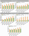
Relative gene expression for matrix formation markers—(a) RUNX2—and (b) COL1A1 and matrix mineralization markers—(c) ALPL, (d) SPP1, and (e) BGLAP. The results are shown as means (n = 4, two technical repetitions), and bars represent SEM. * and # represent statistical analyses between uncoated Ti and tested samples for unstimulated cells and biofilm-stimulated cells, respectively. (*, #, & p < 0.05; **, ##, && p < 0.01; ***, ###, &&& p < 0.001). Ti: titanium alloy Ti6Al4V; WPI: whey protein isolate; PG: phloroglucinol.
Figure 7
Figure 7
Relative gene expression of pro-inflammatory markers: (a) IL1A, (b) IL1B, and (c) IL8. The results are shown as means (n = 4, two technical repetitions), and bars represent SEM. * and # represent significant differences between uncoated Ti and tested samples for unstimulated cells and biofilm-stimulated cells, respectively. (*, # p < 0.05; ## p < 0.01; ###, &&& p < 0.001). Ti: titanium alloy Ti6Al4V; WPI: whey protein isolate; PG: phloroglucinol.

References

    1. Ma Y., Wang S., Wang H., Chen X., Shuai Y., Wang H., Mao Y., He F. Mesenchymal Stem Cells and Dental Implant Osseointegration during Aging: From Mechanisms to Therapy. Stem Cell Res. Ther. 2023;14:382. doi: 10.1186/s13287-023-03611-1. - DOI - PMC - PubMed
    1. Emam S.M., Moussa N. Signaling Pathways of Dental Implants’ Osseointegration: A Narrative Review on Two of the Most Relevant; NF-κB and Wnt Pathways. BDJ Open. 2024;10:29. doi: 10.1038/s41405-024-00211-w. - DOI - PMC - PubMed
    1. Lasserre J.F., Brecx M.C., Toma S. Oral Microbes, Biofilms and Their Role in Periodontal and Peri-Implant Diseases. Materials. 2018;11:1802. doi: 10.3390/ma11101802. - DOI - PMC - PubMed
    1. Seo B.Y., Son K., Son Y.-T., Dahal R.H., Kim S., Kim J., Hwang J., Kwon S.-M., Lee J.-M., Lee K.-B., et al. Influence of Dental Titanium Implants with Different Surface Treatments Using Femtosecond and Nanosecond Lasers on Biofilm Formation. J. Funct. Biomater. 2023;14:297. doi: 10.3390/jfb14060297. - DOI - PMC - PubMed
    1. Kligman S., Ren Z., Chung C.-H., Perillo M.A., Chang Y.-C., Koo H., Zheng Z., Li C. The Impact of Dental Implant Surface Modifications on Osseointegration and Biofilm Formation. J. Clin. Med. 2021;10:1641. doi: 10.3390/jcm10081641. - DOI - PMC - PubMed

LinkOut - more resources