. 2025;49(5):789-811.
doi: 10.32604/biocell.2025.062325. Epub 2025 May 27.

Integrin Alpha8 Beta1 (81): An In-Depth Review of an Overlooked RGD-Binding Receptor

Affiliations

Integrin Alpha8 Beta1 (81): An In-Depth Review of an Overlooked RGD-Binding Receptor

Iman Ezzat et al. Biocell. 2025.

Abstract

Integrins are heterodimeric transmembrane receptors that mediate bidirectional interactions between the intracellular cytoskeletal array and the extracellular matrix. These interactions are critical in tissue development and function by regulating gene expression and sustaining tissue architecture. In humans, the integrin family is composed of 18 alpha (α) and 8 beta (β) subunits, constituting 24 distinct αβ combinations. Based on their structure and ligand-binding properties, only a subset of integrins, 8 out of 24, recognizes the arginine-glycine-aspartate (RGD) tripeptide motif in the native ligand. One of the major RGD binding integrins is integrin alpha 8 beta 1 (α8β1), a central Ras homolog gene family member A (RHOA)-dependent modulator highly expressed in cells with contractile function. This review focuses on the recent advances regarding α8β1 function during organ development, with a particular interest in kidney and inner ear development. We also discuss α8β1's role in injury and disease and its importance for mesenchymal to epithelial transition during cancer development. Finally, we highlight α8β1's importance for hearing function and its future use as a potential diagnostic and therapeutic tool for disease elimination.

Keywords: RGD binding integrin; Transmembrane receptor; cytoskeleton.


Conflict of interest statement

Conflicts of Interest: The authors declare no conflicts of interest to report regarding the present study.

Figures

Figure 1:
Figure 1:
Structure and conformational states of RGD-binding integrins. A: Structural illustration of the RGD-binding integrin α-subunit (left, yellow) and β-subunit (right, blue). B: Conformational states showing transitions from bent closed (low affinity) to extended-open (high affinity), with the ligand-binding pocket interacting with the RGD motif. Biorender.com
Figure 2:
Figure 2:
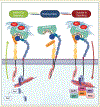
Bidirectional signaling of RGD-binding integrins. The figure illustrates the dynamic transitions of RGD-binding integrins through their resting, inside-out, and outside-in signaling states. In the resting state, integrins adopt a bent-closed conformation with low affinity, stabilized by filamin binding to both α and β subunits’ cytoplasmic tail (CT). Inside-out signaling begins when talin and kindlin bind the β-subunit CT, displacing filamin and inducing a conformational change to an extended-open state (high binding affinity). This process is enhanced by extracellular Mg2+ and mechanical ECM forces. In neutrophils, chemokine-activated Rap1 recruits talin to drive integrin activation. Upon ligand binding, integrins cluster at adhesion sites and initiate outside-in signaling, where filamin repositions to the a-subunit CT, linking integrins to actin filaments. This activates focal adhesion complexes and downstream pathways, including FAK, SRC, AKT, MAPK, ERK, and RHO, regulating adhesion, migration and cytoskeletal reorganization. Arg: arginine, Gly: glycine, Asp: asparagine. Created with Biorender
Figure 3:
Figure 3:
Integrin α8β1 expression and localization in the inner ear and kidney. A–B: α8β1 in the kidney localizes in the glomerular mesangial cells. A: Anti-α8 (red). B: RNAscope (Multiplex v2 Kit, Cat# 323271 ACDBio) for α8 (magenta). Sections were counterstained with DAPI. C–E: α8 in the vestibular system localizes at the cilia (magenta) and stereocilia (data not shown) levels. α8: green, phalloidin (Cat# A12381, ThermoFisher Sci.): red, and acetylated tubulin (Cat# T6793, Sigma-Aldrich): magenta. F–G: α8β1 in the organ of Corti, localizes in the hair cell bundle and cilia. α8β1: green, phalloidin: red and acetylated tubulin: white. Anti-α8 was a gift from Dr. L. Reichardt). Scale bars = A–B, F–G: 10 μm. C–E: 5 μm
Figure 4:
Figure 4:
Illustration of integrin expression and function across organs: this figure represents α8β1 integrin expression and function across various organs. The anatomical diagram is based on human anatomy, with the expression data derived from both in-vitro and in-vivo studies. Created in https://BioRender.com

Similar articles

References

    1. Gravina AN, D’Elia ND, Benedini LA, Messina P. A commentary on the interplay of biomaterials and cell adhesion: new insights in bone tissue regeneration. BIOCELL. 2024;48(11):1517–20. doi: 10.32604/biocell.2024.055513. - DOI
    1. Kadry YA, Calderwood DA. Chapter 22: structural and signaling functions of integrins. Biochim Biophys Acta Biomembr. 2020;1862(5):183206. doi: 10.1016/j.bbamem.2020.183206. - DOI - PMC - PubMed
    1. Wen L, Lee S, Ley K. A splice variant of kindlin-3 is functional in β2 integrin activation and neutrophil adhesion. J Immunol. 2024;212(1_Supplement):1156–5049. doi: 10.4049/jimmunol.212.supp.1156.5049. - DOI
    1. Fagerholm SC, Guenther C, Llort Asens M, Savinko T, Uotila LM. Beta2-integrins and interacting proteins in leukocyte trafficking, immune suppression, and immunodeficiency disease. Front Immunol. 2019;10:254. doi: 10.3389/fimmu.2019.00254. - DOI - PMC - PubMed
    1. Harjunpää H, Llort Asens M, Guenther C, Fagerholm SC. Cell adhesion molecules and their roles and regulation in the immune and tumor microenvironment. Front Immunol. 2019;10:1078. doi: 10.3389/fimmu.2019.01078. - DOI - PMC - PubMed

LinkOut - more resources