Case Reports
. 2025 May 12;17(5):e83986.
doi: 10.7759/cureus.83986. eCollection 2025 May.

An Imitator Within: A Case of Appendiceal Neuroma Masquerading as Chronic Appendicitis

Affiliations
Case Reports

An Imitator Within: A Case of Appendiceal Neuroma Masquerading as Chronic Appendicitis

Zhen Yun Siew et al. Cureus. .

Abstract

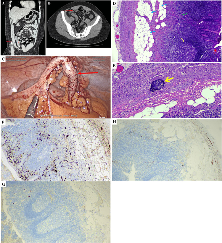
The incidence of appendicitis, an inflammation of the appendix, has been rising significantly in many countries worldwide. Appendicitis may present in various forms, including acute or chronic inflammation, and can manifest as either uncomplicated or complicated conditions, etc. Among these conditions, chronic appendicitis is a rare type of appendiceal inflammation that is less common than acute appendicitis. In this case report, we describe a case of idiopathic appendiceal neuroma, which was initially misdiagnosed as acute gastroenteritis. Diagnosis was further supported by comprehensive laboratory testing, computed tomography and ultrasound imaging, as well as immunohistochemical staining. The patient made a full recovery following a laparoscopic appendectomy. This case highlights appendiceal neuroma as an elusive entity with atypical presentation of appendicitis and chronic histological features, emphasising the critical role of histopathology in establishing a definitive diagnosis.

Keywords: appendicitis; chronic; fibrous obliteration; gastroenteritis; neuroma.


Conflict of interest statement

Human subjects: Consent for treatment and open access publication was obtained or waived by all participants in this study. Conflicts of interest: In compliance with the ICMJE uniform disclosure form, all authors declare the following: Payment/services info: All authors have declared that no financial support was received from any organization for the submitted work. Financial relationships: All authors have declared that they have no financial relationships at present or within the previous three years with any organizations that might have an interest in the submitted work. Other relationships: All authors have declared that there are no other relationships or activities that could appear to have influenced the submitted work.

Figures

Figure 1
Figure 1. CT-scan images (A, B), a picture of the appendix during surgery (C), micrographs of the biopsied appendix stained with haematoxylin and eosin (D, E), and immunohistochemical staining (F-H).
The appendix (red arrow) was identified in the coronal (A) and axial (B) views of the CT-scan image. No focal lesion or mass, no peripancreatic collection, and no streakiness of the adjacent fat were seen. Distended fluid-filled loops of small and large bowel were observed. Thickened distal ileum and caecum were noted. The stomach, small bowel and the rest of the large bowel appeared normal. No obvious bowel-related mass was observed. The appendix was noted to be about 5-6 mm in diameter, and no appendicular mass was observed. (C) Congested blood vessels were observed on the outer surface of the appendix (red arrow), and no perforation can be observed. (D) The cross-sectional view of the appendix (40x magnification) shows fibrosis and fibrofatty tissue infiltration (blue arrows). A reactive lymphoid follicle (gold arrow) was observed surrounded by diffuse lymphoid tissue. Diffuse lymphoid tissue was observed with the loss of the germinal centre (green arrow). Chronic inflammatory cell infiltration (white arrow) was observed in the inner region of a crypt. A mucosal erosion (red open arrow) was noted. Scattered neutrophils were present within the crypt epithelium and mucosa. The wall is infiltrated by mild lymphocytes. Serosa was fibrotic and had no evidence of parasites, granuloma or malignancy. (E) Under 100x magnification, a lymphoid aggregate (yellow open arrow) was observed somewhere in between the submucosa and muscularis layers of the appendix. Immunohistochemical (IHC) staining was performed to stain S-100 protein (F), chromogranin A (G), and synaptophysin (H). Micrographs F-H were captured at 40x magnification.

Similar articles

References

    1. The global, regional, and national burden of appendicitis in 204 countries and territories, 1990-2019: a systematic analysis from the Global Burden of Disease Study 2019. Guan L, Liu Z, Pan G, Zhang B, Wu Y, Gan T, Ouyang G. BMC Gastroenterol. 2023;23:44. - PMC - PubMed
    1. Effect of the Movement Control Order on the Incidence of Complicated Appendicitis During the COVID-19 Pandemic: A Cross-Sectional Study. Sukiman H, Mohamad AM, Raduan MF, Yasim MN, Lazim MI. Malays J Med Sci. 2021;28:130–136. - PMC - PubMed
    1. A Study on the Clinical Factors Associated with Acute Appendicitis and Perforated Appendicitis among Children in a Secondary Medical Centre in Malaysia. Tan PH, Teng XX, Gan ZY, Tan SQ. Malays J Med Sci. 2020;27:139–146. - PMC - PubMed
    1. Chronic Appendicitis, the Lesser-Known Form of Appendiceal Inflammation: A Case Report. Lee CK, Pelenyi SS, Fleites O, Velez V, Alaimo KL, Ramcharan DN, Tiesenga F. Cureus. 2021;13:0. - PMC - PubMed
    1. Typical and Atypical Presentations of Appendicitis and Their Implications for Diagnosis and Treatment: A Literature Review. Echevarria S, Rauf F, Hussain N, et al. Cureus. 2023;15:0. - PMC - PubMed

Publication types

LinkOut - more resources