. 2025 May 29:16:1562116.
doi: 10.3389/fphar.2025.1562116. eCollection 2025.

Therapeutic potential of Cordyceps militaris cultivated with Ginkgo biloba seeds for alleviating western diet-induced type 2 diabetes and diabetic nephropathy

Affiliations

Therapeutic potential of Cordyceps militaris cultivated with Ginkgo biloba seeds for alleviating western diet-induced type 2 diabetes and diabetic nephropathy

Shinn-Zong Lin et al. Front Pharmacol. .

Abstract

Background: Diabetic nephropathy (DN), a leading cause of chronic kidney disease and end-stage renal disease, is a serious complication of type 2 diabetes mellitus (T2DM). Current therapies primarily slow disease progression but are unable to reverse kidney damage, highlighting the need for novel therapy to treat DN.

Objective: This study evaluated the therapeutic potential of Cordyceps militaris (C. militaris) cultivated on Ginkgo biloba (G. biloba) seeds in ameliorating T2DM and its complications, especially DN. A T2DM mouse model was established using ApoE knockout mice fed a Western diet (WD).

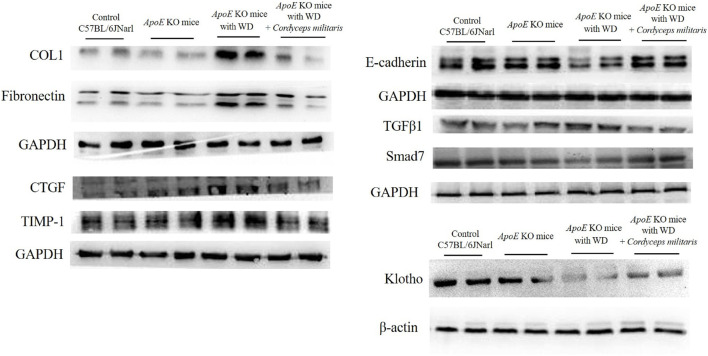
Results: Treatment with the specially cultivated C. militaris ameliorated hyperglycemia, dyslipidemia and hepatic dysfunction, while mitigating T2DM-induced renal damage. Key biochemical markers, including blood glucose, triglycerides, cholesterol, blood urea nitrogen (BUN), and creatinine, were significantly improved after treatment. Histopathologic analysis revealed restored renal morphology, reduced fibrosis and decreased amyloid deposition. Mechanistic studies showed downregulation of fibrosis-related proteins such as α-SMA, COL1, TIMP-1, CTGF, TGFβ1 and fibronectin, and upregulation of E-cadherin, Smad7 and Klotho, proteins with anti-fibrotic and renoprotective properties.

Conclusion: These results suggest that the specially cultivated C. militaris enhances metabolic regulation and renal repair mechanisms, effectively attenuating T2DM-induced renal damage. This unique cultivation approach enriches the bioactive properties of C. militaris and offers a promising natural therapeutic strategy for T2DM and DN. Further studies are needed to validate these results in clinical settings and to explore long-term efficacy and safety.

Keywords: Cordyceps militaris; Ginkgo biloba seeds; diabetes mellitus; diabetic nephropathy; kidney.


Conflict of interest statement

Author S-JC was employed by Home Run Biotechnology Co., Ltd. The remaining authors declare that the research was conducted in the absence of any commercial or financial relationships that could be construed as a potential conflict of interest.

Figures

FIGURE 1
FIGURE 1
Changes in body weight of ApoE KO mice under different experimental conditions. The group of ApoE KO mice fed a Western diet (WD) without treatment showed an initial increase in body weight followed by a decrease after 14 weeks. In contrast, ApoE KO mice fed a WD and treated with this specially cultivated Cordyceps militaris from week 8 maintained a stable body weight throughout the experimental period. Data are expressed as mean ± SD. *** = p < 0.001 compared to ApoE KO mice fed a standard diet group; ## = p < 0.01 compared to ApoE KO mice fed with WD without any treatment.
FIGURE 2
FIGURE 2
Serum glucose levels in ApoE KO mice under different experimental conditions. The ApoE KO mice fed a Western diet (WD) alone showed a significant increase in serum glucose levels by week 8. After initiation of this specially cultivated Cordyceps militaris treatment at week 8, a progressive decrease in serum glucose levels was observed, which continued consistently until week 16. Data were expressed as mean ± SD. *** = p < 0.001 compared to ApoE KO mice fed a standard diet group; ### = p < 0.001, # = p < 0.05 compared to ApoE KO mice fed with WD without any treatment.
FIGURE 3
FIGURE 3
Biochemical analysis of blood lipid levels and liver function in ApoE KO mice under different experimental conditions. (A) The ApoE KO mice fed a Western diet (WD) showed significantly elevated levels of triglycerides and total cholesterol, which were reduced after this specially cultivated Cordyceps militaris treatment. (B) Elevated serum concentrations of aspartate aminotransferase (AST) and alanine aminotransferase (ALT), markers of liver dysfunction induced by WD, were significantly reduced in the Cordyceps militaris-treated group. Data were expressed as mean ± SD. *** = p < 0.001 compared to ApoE KO mice fed a standard diet group; ### = p < 0.001 compared to ApoE KO mice fed with WD without any treatment.
FIGURE 4
FIGURE 4
Evaluation of renal dysfunction and histopathologic changes in ApoE KO mice under different experimental conditions. (A) The ApoE KO mice fed a Western diet (WD) showed changes in renal morphology and a decrease in renal weight. (B) The ApoE KO mice showed significantly elevated blood urea nitrogen (BUN) and serum creatinine levels, indicating renal dysfunction. However, this specially cultivated Cordyceps militaris treatment improved the kidney and significantly decreased the BUN and serum creatinine levels. Data were expressed as mean ± SD. *** = P < 0.001 compared to ApoE KO mice fed a standard diet group; ### = P < 0.001 compared to ApoE KO mice with WD without any treatment.
FIGURE 5
FIGURE 5
Restoration of renal glomerular morphology by Cordyceps militaris treatment in ApoE KO mice. Histopathological analysis using Hematoxylin and eosin staining showed severe morphological abnormalities in renal glomeruli, including shrinkage and atrophy, in WD-fed mice. These pathological changes were ameliorated in the Cordyceps militaris-treated group and the glomerular morphology resembled that of healthy controls.
FIGURE 6
FIGURE 6
Evaluation of collagen deposition in renal tissue using histological staining. (A) Masson’s trichrome staining and (B) Picrosirius red staining revealed significant collagen accumulation in the kidneys of ApoE KO mice fed a Western diet (WD). This specially cultivated Cordyceps militaris treatment remarkably reduced collagen deposition as evidenced by reduced staining intensity and improved tissue morphology.
FIGURE 7
FIGURE 7
Analysis of fibrosis and amyloid deposition in the kidneys of ApoE KO mice. (A) Immunohistochemical staining revealed significantly increased expression of α-smooth muscle actin (α-SMA), a marker of fibrosis, in the kidneys of mice fed a Western diet (WD). This specially cultivated Cordyceps militaris treatment significantly reduced α-SMA expression, demonstrating its antifibrotic effects. (B) Toluidine blue staining showed extensive amyloid deposits in the renal glomeruli of WD-fed mice, which were attenuated after this specially cultivated Cordyceps militaris administration, indicating its protective role against amyloid accumulation.
FIGURE 8
FIGURE 8
Western blot analysis of fibrosis-related proteins and Klotho expression in the kidneys of ApoE KO mice. Western diet (WD)-fed mice showed upregulation of fibrosis-associated proteins, including collagen type I (COL1), fibronectin, connective tissue growth factor (CTGF), tissue inhibitor of metalloproteinase 1 (TIMP-1), and transforming growth factor β1 (TGFβ1), and downregulation of anti-fibrotic proteins E-cadherin and Smad7. This specially cultivated Cordyceps militaris treatment reversed these trends by reducing pro-fibrotic proteins, restoring E-cadherin and Smad7 expression, and increasing Klotho level.

Similar articles

References

    1. Al-Jameil N., Khan F. A., Arjumand S., Khan M. F., Tabassum H. (2014). Associated liver enzymes with hyperlipidemic profile in type 2 diabetes patients. Int. J. Clin. Exp. Pathol. 7, 4345–4349. - PMC - PubMed
    1. Beckman J. A., Creager M. A. (2016). Vascular complications of diabetes. Circ. Res. 118, 1771–1785. 10.1161/CIRCRESAHA.115.306884 - DOI - PubMed
    1. Belwal T., Giri L., Bahukhandi A., Tariq M., Kewlani P., Bhatt I. D., et al. (2019). “Ginkgo biloba,” in Nonvitamin and nonmineral nutritional supplements. New York, NY, United States, Elsevier, 241–250.
    1. Biernacka P., Adamska I., Felisiak K. (2023). The potential of ginkgo biloba as a source of biologically active compounds-A review of the recent literature and patents. Molecules 28, 3993. 10.3390/molecules28103993 - DOI - PMC - PubMed
    1. Boateng I. D., Yang X. M. (2021). Effect of different drying methods on product quality, bioactive and toxic components of Ginkgo biloba L. seed. J. Sci. Food Agric. 101, 3290–3297. 10.1002/jsfa.10958 - DOI - PubMed

LinkOut - more resources