Review
. 2025 May 29:16:1546041.
doi: 10.3389/fphar.2025.1546041. eCollection 2025.

Targeting programmed cell death with natural products: a potential therapeutic strategy for diminished ovarian reserve and fertility preservation

Affiliations
Review

Targeting programmed cell death with natural products: a potential therapeutic strategy for diminished ovarian reserve and fertility preservation

Wenhan Ju et al. Front Pharmacol. .

Abstract

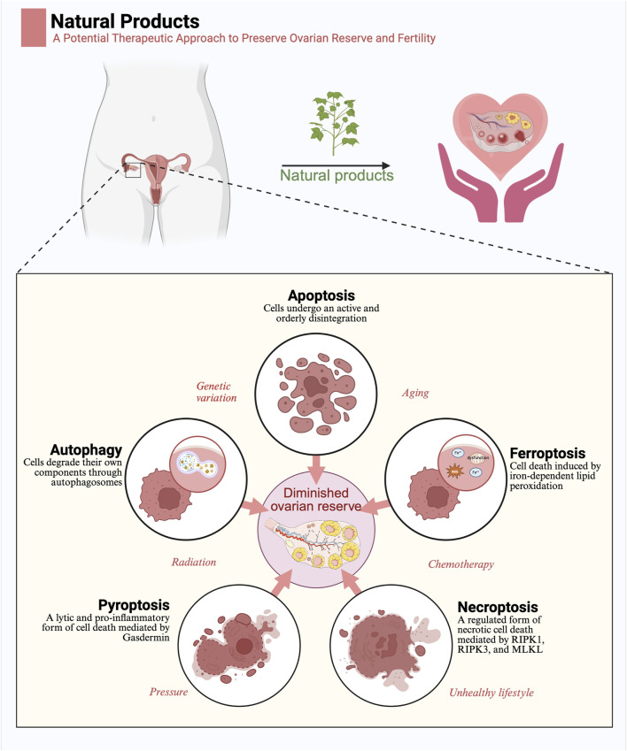
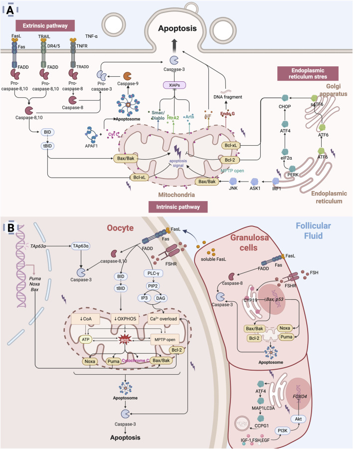
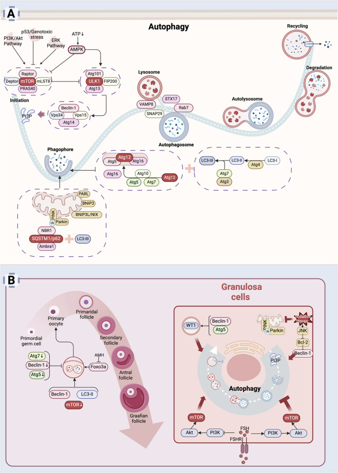
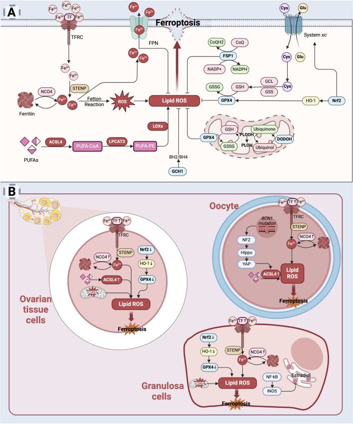
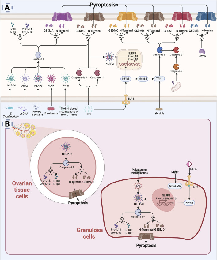
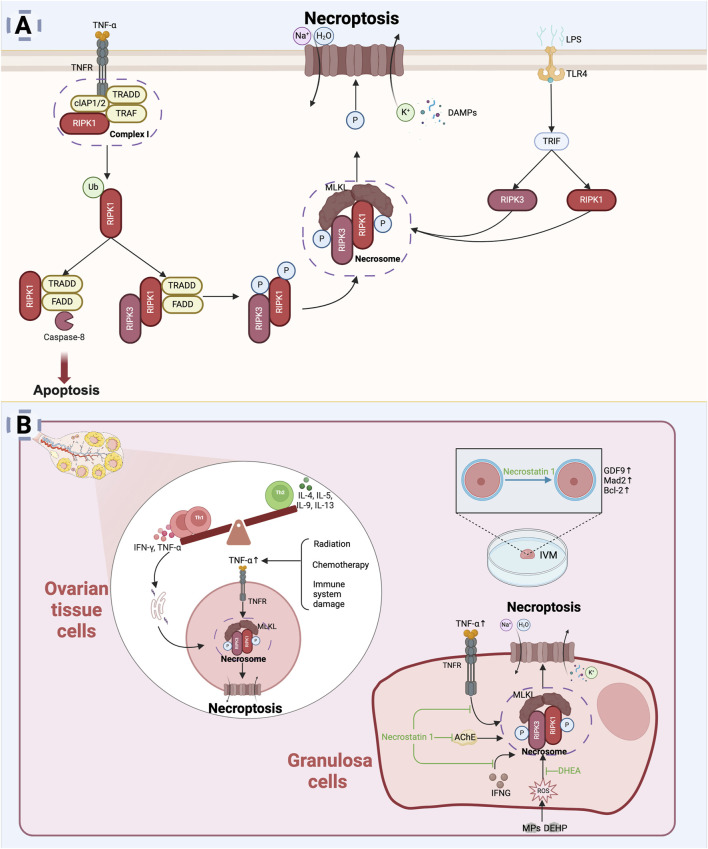
The depletion of ovarian reserve is a major factor contributing to the decline in female fertility. It is characterized by a simultaneous reduction in the quantity and quality of oocytes and the follicular pools. The cyclic recruitment of primordial follicles and the preservation of oocyte quality involve complex and tightly regulated biological processes. Granulosa cells, which surround the oocytes, play a pivotal role in follicular development and the determination of follicular fate. Programmed cell death (PCD), a genetically regulated process of cell elimination, is a key factor in the regulation of ovarian reserve dynamics. Emerging evidence suggests that natural products derived from medicinal plants, dietary components, animals, and microorganisms may modulate PCD in granulosa cells through various molecular mechanisms and signaling pathways. These natural products have demonstrated preliminary effects in delaying ovarian aging and preserving ovarian reserve in preclinical models. This review discusses the roles and underlying mechanisms of various forms of PCD in diminished ovarian reserve, while summarizing the current findings on natural products that influence granulosa cells PCD to protect ovarian function. These insights may contribute to the future development of novel, targeted strategies aimed at preserving female reproductive potential.

Keywords: IVF; diminished ovarian reserve; female fertility; natural products; programmed cell death.


Conflict of interest statement

The authors declare that the research was conducted in the absence of any commercial or financial relationships that could be construed as a potential conflict of interest.

Figures

FIGURE 1
FIGURE 1
Natural products exert a protective function in diminished ovarian reserve by regulating programmed cell death: Five modes of programmed cell death, including apoptosis, autophagy, ferroptosis, pyroptosis, and necroptosis, are important pathogenetic mechanisms of diminished ovarian reserve. Factors such as genetic mutations, aging, radiation, chemotherapy, stress, and unhealthy lifestyle contribute to diminished ovarian reserve by influencing programmed cell death. Natural products may contribute to the preservation of ovarian reserve by regulating programmed cell death (Created in BioRender.com).
FIGURE 2
FIGURE 2
Apoptosis leads to diminished ovarian reserve: (A) Apoptosis is primarily mediated through three key pathways: the extrinsic pathway (death receptor pathway), the intrinsic pathway (mitochondrial pathway), and the endoplasmic reticulum stress (ER) pathway. The extrinsic pathway is initiated by signals from death receptors on the cell surface, which directly activate caspases, thereby initiating the apoptotic process (Galluzzi et al., 2012; Shakibaei et al., 2005). The intrinsic pathway is typically activated by intracellular stressors such as oxidative stress or DNA damage (Kulikov et al., 2012). This pathway involves mitochondrial alterations, including the release of Cyt C, which activates downstream caspases to induce apoptosis. The ER stress pathway is triggered by dysfunction in the endoplasmic reticulum, leading to increased expression of intracellular pro-apoptotic factors (Huang S. et al., 2019; Liang et al., 2020). (B) Mechanisms of Apoptosis in diminished ovarian reserve. In the context of DOR, oocytes are lost through apoptosis, a process regulated by specific gene expression (Huang et al., 2023). Both the extrinsic and intrinsic apoptotic pathways can be activated to initiate oocyte apoptosis (Zhu et al., 2016). Granulosa cells apoptosis, which disrupts the supportive microenvironment of the oocyte, further accelerates oocyte apoptosis (Zhu et al., 2016; Zhu et al., 2015). Multiple apoptotic pathways, including extrinsic, intrinsic, and ER stress-mediated signaling, contribute to granulosa cell apoptosis and ultimately accelerate ovarian reserve depletion (Inoue et al., 2007; Matsuda et al., 2008; Hsu et al., 1996; Ratts et al., 1995; Kugu et al., 1998; Perez et al., 1999; Li H. et al., 2023) (Created in BioRender.com).
FIGURE 3
FIGURE 3
Autophagy leads to diminished ovarian reserve: (A) Mechanism of autophagy. Autophagy is a process by which cells degrade themselves. Signals such as AMPK, PI3K/Akt, ERK, p53, etc. influence autophagy by regulating mTOR activity (Yang and Klionsky, 2010). After autophagy is initiated, autophagosomes are assembled inside the cell to wrap damaged organelles or aggregated proteins inside the cell (Mizushima and Komatsu, 2011). Subsequently, autophagosomes fuse with lysosomes to form autophagic lysosomes, and the internal material is degraded by lysosomal enzymes (Itakura et al., 2012). Finally, the degradation products are released back into the cytoplasm for re-synthesis of substances needed by the cell or to provide energy. (B) Mechanism of cellular autophagy in ovarian reserve hypoplasia. Autophagy is involved in the maintenance of the primordial follicle number (Cao et al., 2017; Sonigo et al., 2019). Autophagy helps granulosa cells adapt to adverse external stimuli and promotes granulosa cells survival (Shao et al., 2022). Excessive autophagy also induces granulosa cells death (Cao et al., 2018) (Created in BioRender.com).
FIGURE 4
FIGURE 4
Ferroptosis and its role in diminished ovarian reserve: (A) Mechanism of ferroptosis: Excessive intracellular iron accumulation promotes the generation of free radicals, particularly lipid peroxides (Jiang et al., 2021). Glutathione peroxidase 4 (GPX4) plays a critical role in preventing lipid peroxidation; however, the inhibition of GPX4 activity during ferroptosis exacerbates lipid peroxidation (Liang et al., 2022; Xie et al., 2023). The accumulation of lipid peroxides causes structural damage to the cellular membrane, ultimately resulting in cell death. (B) Mechanism of cellular ferroptosis in diminished ovarian reserve: In the ovary, iron accumulation may increase due to aging, oxidative stress, or pathological conditions (Sze et al., 2022; Zhang et al., 2018). This excess iron triggers oxidative stress, leading to damage in ovarian cells, including oocytes and granulosa cells (Wang F. et al., 2022). Concurrently, reduced levels of glutathione (GSH) and inhibition of GPX4 activity weaken the antioxidant defense system, further promoting ferroptosis (Niu et al., 2023; Zhang S. et al., 2023). This process contributes to cellular damage and diminished ovarian reserve (Created in BioRender.com).
FIGURE 5
FIGURE 5
Pyroptosis leading to diminished ovarian reserve: (A) Mechanism of pyroptosis. Pyroptosis is initiated by the activation of inflammasomes (e.g., NLRP3, AIM2) in response to infection or cellular injury (Frank and Vince, 2019). Activated inflammasomes contribute to the activation of proteases such as caspase-1. Caspase-1 cleaves the N-terminal structural domain of the GSDMD to insert into the cell membrane, forming a pore that leads to rupture of the membrane and release of the cellular contents (Kovacs and Miao, 2017). At the same time, caspase-1 promotes the processing, maturation and release of IL-1β and IL-18 (Kovacs and Miao, 2017). LPS can directly activate Caspase-4/5 (human) or Caspase-11 (mouse) in host cells to cleave GSDMD (Sahoo et al., 2023). Caspase-8, Caspase-3, and GzmA are also involved in GSDM protein cleavage leading to cellular pyroptosis (Orning et al., 2018; Zhang JY. et al., 2021; Wang et al., 2021; Wang et al., 2017; Zhou et al., 2020). (B) Mechanisms of pyroptosis in diminished ovarian reserve. In the ovary, environmental pollutants, oxidative stress, or chronic inflammation can activate inflammasomes (e.g., NLRP3), leading to intracellular signaling (Navarro-Pando et al., 2021; Lliberos et al., 2021b; Khallaf et al., 2023; Chi et al., 2023; Liu K. et al., 2024; Xie et al., 2024). Pyroptosis not only directly affects the survival of granulosa cells, but may also lead to changes in the ovarian microenvironment, affecting ovarian reserve function (Zhou C. et al., 2024) (Created in BioRender.com).
FIGURE 6
FIGURE 6
Necroptosis and diminished ovarian reserve. (A) Mechanism of necroptosis: TNF-α and its receptor TNFR mediate necroptosis (Nailwal and Chan, 2019), with RIPK1 phosphorylation activating RIPK3 to form the necrosome (Nailwal and Chan, 2019). RIPK3 phosphorylation activates MLKL, which translocates to the cell membrane, causing membrane rupture, leakage of cellular contents, and cell death (Murao et al., 2021). TLR4 can also induce necroptosis through TRIF interaction with the necrosome (Solon et al., 2024). (B) Mechanisms of necroptosis in diminished ovarian reserve: In the ovarian microenvironment, chronic inflammation or harmful stimuli may trigger TNF-α release, activating necroptotic pathways (Khallaf et al., 2023; Li X. et al., 2023; Akdemir et al., 2022; Tao et al., 2024). Necrostatin-1 promotes granulosa cells survival and improves IVM oocyte quality by inhibiting necroptosis (Hojo et al., 2016; Jo et al., 2015) (Created in BioRender.com).
FIGURE 7
FIGURE 7
Meta analysis results of natural product treatment for DOR. (A) Risk bias analysis; (B) Forest Plot of live birth rate; (C) Forest Plot of clinical pregnancy rate.

Similar articles

References

    1. Agwuegbo U. T., Colley E., Albert A. P., Butnev V. Y., Bousfield G. R., Jonas K. C. (2021). Differential FSH glycosylation modulates FSHR oligomerization and subsequent cAMP signaling. Front. Endocrinol. 12, 765727. 10.3389/fendo.2021.765727 - DOI - PMC - PubMed
    1. Akdemir Y., Akpolat M., Elmas O., Kececi M., Buyukuysal C., Cetinkaya B., et al. (2022). Capsaicin prevents radiotherapy-induced premature ovarian failure in rats. Reprod. Fertil. Dev. 34 (3), 350–361. 10.1071/RD21235 - DOI - PubMed
    1. Ashkenazi A., Dixit V. M. (1998). Death receptors: signaling and modulation. Science 281 (5381), 1305–1308. 10.1126/science.281.5381.1305 - DOI - PubMed
    1. Baddela V. S., Sharma A., Vanselow J. (2020). Non-esterified fatty acids in the ovary: friends or foes? Reprod. Biol. Endocrinol. 18, 60. 10.1186/s12958-020-00617-9 - DOI - PMC - PubMed
    1. Bagavant H., Adams S., Terranova P., Chang A., Kraemer F. W., Lou Y., et al. (1999). Autoimmune ovarian inflammation triggered by proinflammatory (Th1) T cells is compatible with normal ovarian function in mice. Biol. Reprod. 61 (3), 635–642. 10.1095/biolreprod61.3.635 - DOI - PubMed

LinkOut - more resources