. 2024 Jan 4;11(1):2300564.
doi: 10.1002/admi.202300564. Epub 2023 Oct 15.

Interaction of Blood and Bacteria with Slippery Hydrophilic Surfaces

Affiliations

Interaction of Blood and Bacteria with Slippery Hydrophilic Surfaces

Prem Kantam et al. Adv Mater Interfaces. .

Abstract

Slippery surfaces (i.e., surfaces that display high liquid droplet mobility) are receiving significant attention due to their biofluidic applications. Non-textured, all-solid, slippery hydrophilic (SLIC) surfaces are an emerging class of rare and counter-intuitive surfaces. In this work, the interactions of blood and bacteria with SLIC surfaces are investigated. The SLIC surfaces demonstrate significantly lower platelet and leukocyte adhesion (≈97.2% decrease in surface coverage), and correspondingly low platelet activation, as well as significantly lower bacterial adhesion (≈99.7% decrease in surface coverage of live Escherichia Coli and ≈99.6% decrease in surface coverage of live Staphylococcus Aureus) and proliferation compared to untreated silicon substrates, indicating their potential for practical biomedical applications. The study envisions that the SLIC surfaces will pave the path to improved biomedical devices with favorable blood and bacteria interactions.

Keywords: bacterial adhesion; hydrophilicity; platelet activation; platelet adhesion; slippery.


Conflict of interest statement

Conflict of Interest The authors declare no conflict of interest.

Figures

Figure 1.
Figure 1.
Characterization of SLIC surfaces. A) High-resolution C1s XPS spectra of untreated silicon and SLIC surfaces. The C─C peak on untreated silicon is due to the presence of adventitious carbon. The C─O peak on SLIC surface indicates the presence of PEG functional groups. B) AFM image depicting the topography of SLIC surface with low surface roughness (Rrms<1nm). C) Time-lapse images of a 20 μL droplet adhered (i.e., not sliding) on an untreated silicon surface at a tilt angle of 8°. D) Time-lapse images of a 20 μL droplet sliding on a SLIC surface at a tilt angle of 8°.
Figure 2.
Figure 2.
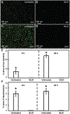
Platelet & leukocyte adhesion on SLIC surfaces. A,B) Fluorescent microscopy images showing blood cell (live) adhesion (stained green) on untreated silicon and SLIC surfaces, respectively. C) Bar chart indicating significant surface coverage (*indicates p < 0.05) of live blood cells on untreated silicon and SLIC surfaces. D,E) Fluorescent microscopy images showing platelet & leukocyte adhesion (stained red) on untreated silicon and SLIC surfaces, respectively. F) Bar chart indicating significant surface coverage (*indicates p < 0.05) of platelets & leukocytes (combined) on untreated silicon and SLIC surfaces. G,H) Fluorescent microscopy images showing leukocyte adhesion (stained blue) on untreated silicon and SLIC surfaces, respectively. I) Bar chart indicating significant surface coverage (*indicates p < 0.05) of leukocytes on untreated silicon and SLIC surfaces.
Figure 3.
Figure 3.
Platelet activation and cytotoxicity on SLIC surfaces. A,B) SEM images showing platelet activation on untreated silicon and SLIC surfaces, respectively at lower magnification. C,D) SEM images showing platelet activation on untreated silicon and SLIC surfaces, respectively at higher magnification. E) Bar chart comparing the cytotoxicity on negative control, positive control, untreated silicon and SLIC surfaces (*indicates p < 0.05).
Figure 4.
Figure 4.
E. coli adhesion on SLIC surfaces using fluorescence microscopy. A,B) Fluorescent microscopy images showing E. coli adhesion on untreated silicon and SLIC surfaces, respectively after 6 h of incubation. C,D) Fluorescent microscopy images showing E. coli adhesion on untreated silicon and SLIC surfaces, respectively after 24 h of incubation. E,F) Bar charts indicating the significant surface coverage (*indicates p < 0.05) of live and dead E. coli, respectively on untreated silicon compared to SLIC surfaces at 6 and 24 h of incubation. The area fraction of bacteria adhesion was ≈0.1% on our SLIC surfaces, and there was no discernible change at 24 h compared to 6 h.
Figure 5.
Figure 5.
E. coli adhesion on SLIC surfaces using SEM. A–D) SEM images showing E. coli adhesion on untreated silicon and SLIC surfaces, respectively after 6 h of incubation. E–H) SEM images showing E. coli adhesion on untreated silicon and SLIC surfaces, respectively after 24 h of incubation.
Figure 6.
Figure 6.
S. aureus adhesion on SLIC surfaces using fluorescence microscopy. A,B) Fluorescent microscopy images showing S. aureus adhesion on untreated silicon and SLIC surfaces, respectively after 6 h of incubation. C,D) Fluorescent microscopy images showing S. aureus adhesion on untreated silicon and SLIC surfaces, respectively after 24 h of incubation. E,F) Bar charts indicating the significant surface coverage (*indicates p < 0.05) of live and dead S. aureus, respectively on untreated silicon compared to SLIC surfaces at 6 and 24 h of incubation. The area fraction of bacteria adhesion was ≈0.1% on our SLIC surfaces, and there was no discernible change at 24 h compared to 6 h.
Figure 7.
Figure 7.
S. aureus adhesion on SLIC surfaces using SEM. A–D) SEM images showing S. aureus adhesion on untreated silicon and SLIC surfaces, respectively after 6 h of incubation. E–H) SEM images showing S. aureus adhesion on untreated silicon and SLIC surfaces, respectively after 24 h of incubation.

Similar articles

References

    1. Movafaghi S, Wang W, Bark DL, Dasi LP, Popat KC, Kota AK, Mater. Horiz 2019, 6, 1596; - PMC - PubMed
    2. Zhang P, Lv FY, Energy 2015, 82, 1068;
    3. Li J, Ueda E, Paulssen D, Levkin PA, Adv. Funct. Mater 2019, 29, 1802317;
    4. Cha H, Vahabi H, Wu A, Chavan S, Kim M-K, Sett S, Bosch SA, Wang W, Kota AK, Miljkovic N, Sci. Adv 2020, 6, eaax0746; - PMC - PubMed
    5. Lee C, Choi C-H, Kim C-J, Exp. Fluids 2016, 57, 176;
    6. Wang W, Sun J, Vallabhuneni S, Pawlowski B, Vahabi H, Nellenbach K, Brown AC, Scholle F, Zhao J, Kota AK, Mater. Horiz 2022, 9, 2863; - PMC - PubMed
    7. Channon RB, Menger RF, Wang W, Carrão DB, Vallabhuneni S, Kota AK, Henry CS, Microfluid. Nanofluid 2021, 25, 31.
    1. Vallabhuneni S, Movafaghi S, Wang W, Kota AK, Macromol. Mater. Eng 2018, 303, 1800313;
    2. Tuteja A, Choi W, Ma M, Mabry JM, Mazzella SA, Rutledge GC, Mckinley GH, Cohen RE, Science 2007, 318, 1618; - PubMed
    3. Sutherland DJ, Rather AM, Sabino RM, Vallabhuneni S, Wang W, Popat KC, Kota AK, ACS Sustainable Chem. Eng 2023, 11, 2397; - PMC - PubMed
    4. Epstein AK, Wong T-S, Belisle RA, Boggs EM, Aizenberg J, Proc. Natl. Acad. Sci. U.S.A 2012, 109, 13182; - PMC - PubMed
    5. Movafaghi S, Cackovic MD, Wang W, Vahabi H, Pendurthi A, Henry CS, Kota AK, Adv. Mater. Interfaces 2019, 6, 1900232; - PMC - PubMed
    6. Vahabi H, Wang W, Popat KC, Kwon G, Holland TB, Kota AK, Appl. Phys. Lett 2017, 110, 251602.
    1. Tian X, Verho T, Ras RHA, Science 2016, 352, 142; - PubMed
    2. Peppou-Chapman S, Hong JK, Waterhouse A, Neto C, Chem. Soc. Rev 2020, 49, 3688; - PubMed
    3. Hoque MJ, Sett S, Yan X, Liu D, Rabbi KF, Qiu H, Qureshi M, Barac G, Bolton L, Miljkovic N, ACS Appl. Mater. Interfaces 2022, 14, 4598; - PubMed
    4. Wang W, Vahabi H, Movafaghi S, Kota AK, Adv. Mater. Interfaces 2019, 6, 1900538. - PMC - PubMed
    1. Buddingh JV, Hozumi A, Liu G, Prog. Polym. Sci 2021, 123, 101468;
    2. Wang L, Mccarthy TJ, Angew. Chem., Int. Ed 2016, 55, 244. - PubMed
    1. Wang X, Wang Z, Heng L, Jiang L, Adv. Funct. Mater 2020, 30, 1902686;
    2. Zhao H, Deshpande CA, Li L, Yan X, Hoque MJ, Kuntumalla G, Rajagopal MC, Chang HC, Meng Y, Sundar S, ACS Appl. Mater. Interfaces 2020, 12, 12054; - PubMed
    3. Zhao H, Khodakarami S, Deshpande CA, Ma J, Wu Q, Sett S, Miljkovic N, ACS Appl. Mater. Interfaces 2021, 13, 38666. - PubMed

LinkOut - more resources