. 2025 May 14;36(2):102556.
doi: 10.1016/j.omtn.2025.102556. eCollection 2025 Jun 10.

CRISPR antiviral inhibits neurotrophic JC polyomavirus in 2D and 3D culture models through dual-gRNA excision by SaCas9

Affiliations

CRISPR antiviral inhibits neurotrophic JC polyomavirus in 2D and 3D culture models through dual-gRNA excision by SaCas9

Angela Rocchi et al. Mol Ther Nucleic Acids. .

Abstract

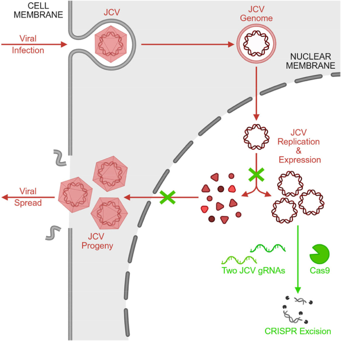
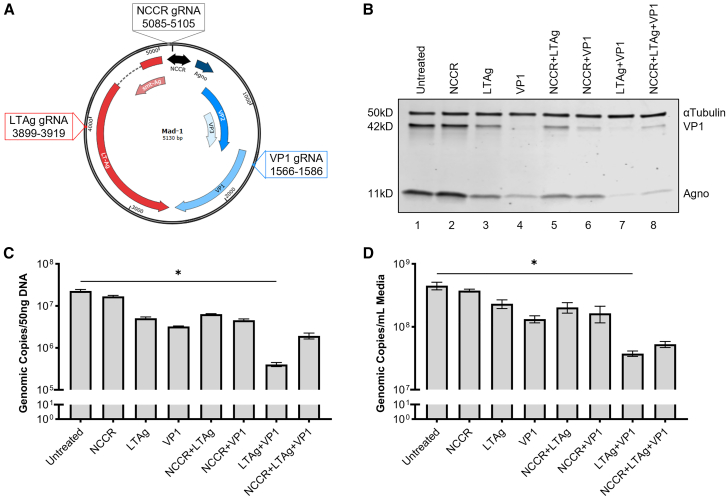
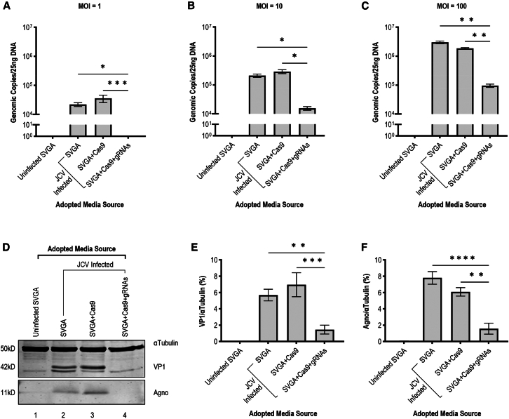
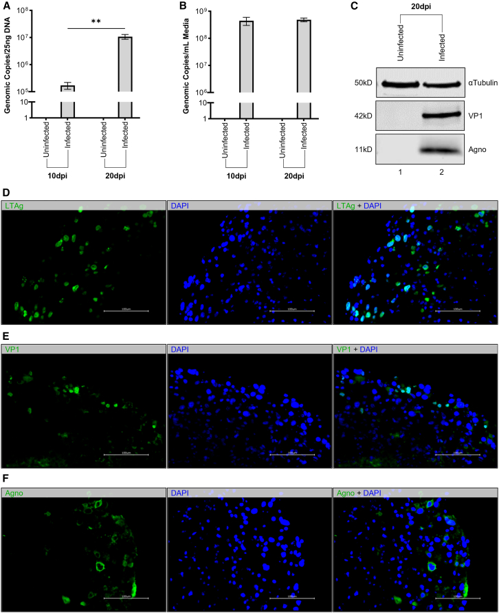
Without an effective antiviral, JC virus (JCV) has persisted throughout multiple epochs of immunosuppression, causing the opportunistic demyelinating disease, progressive multifocal leukoencephalopathy (PML). This study proposes a novel therapy using a dual-gRNA, SaCas9, CRISPR antiviral targeting JCV transcription factor, large tumor antigen (LT-Ag), and capsid protein, viral protein 1 (VP1). This treatment was validated using traditional two-dimensional cell culture. A recombinant cell line was produced from SVG astrocytes (SVGA) via lentiviral inoculation and puromycin selection. Following infection, sanger sequencing identified uniform excision of the circular dsDNA genome of JCV, significantly reducing viral load per genomic copy number on qPCR, viral proteins on western blot, and infectivity of viral progeny on adoptive transfer. Following this proof-of-concept using cell lines, translatability of results was advanced using three-dimensional, heterogeneous cerebral organoids (COs). COs were infected and treated with the lentivirus-packaged CRISPR antiviral. As observed in monolayer culture, a truncated genome was confirmed with sequencing, reducing viral load per genomic copy number on qPCR, protein levels on immunofluorescent imaging, and infectivity on adoptive transfer. The high efficacy of this JCV-targeting CRISPR antiviral in the context of cerebral organoids expounds on its value for the currently untreatable JCV and PML.

Keywords: CRISPR; JC virus; MT: RNA/DNA Editing; antiviral; cerebral organoids; gene editing; progressive multifocal leukoencephalopathy.


Conflict of interest statement

A.R., S.L., C.C., H.L., S.C., A.B., I.K.S., and H.S.W. declare no conflict of interest. K.K. is named inventor on patents that cover the viral gene editing technology that is the subject of this article. K.K. is a co-founder, board member, scientific advisor, and holds equity in Excision BioTherapeutics, a biotech startup that has licensed the viral gene editing technology from Temple University for commercial development and clinical trials.

Figures

None
Graphical abstract
Figure 1
Figure 1
Dual-gRNA SaCas9 construct targeting JCV LT-Ag and VP1 produces the greatest reduction in JCV viral load (A) Map depicting optimal SaCas9-compatible gRNA targets on circular dsDNA genome of Mad-1 per bioinformatic software Benchling. Targeted regions include the NCCR (black), early transcript LT-Ag (red), and late transcript VP1 (blue). Seven separate combinations of these gRNAs were incorporated into SaCas9-construct, pPapi, to determine the most effective antiviral. (B) Western blot depicting viral protein levels of cells treated with CRISPR constructs described above. Compared to untreated control, the largest reduction in both VP1 (42 kD) and Agno (11 kD) is evident for cells treated with the two-gRNA construct targeting LT-Ag and VP1 (LTAg+VP1). (C) Viral genomic copy number measured by qPCR of DNA extracted from cell lysates of conditions described above reveals a statistically significant decrease in cells expressing the LTAg+VP1 dual-gRNA construct compared to untreated control (∗ = p = 0.0229, n = 16). (D) Viral genomic copy number measured by qPCR of DNA extracted from media of cell conditions described above reveals a statistically significant decrease in the media of the LTAg+VP1 dual-gRNA cells compared to untreated control (∗p = 0.0318, n = 16).
Figure 2
Figure 2
Excision of JCV genome linking LT-Ag and VP1 cut sites by dual-gRNA CRISPR construct reduces viral genomic copy number of cells and media and eliminates viral protein expression (A) Illustration of ePCR amplicons produced by primers bound to full-length (“Intact”) and CRISPR-excised (“Remnant”) Mad-1. A full-length genome produces a 2,919 bp amplicon; following excision of the region linking gRNA cut sites, a 583 bp amplicon (green) is produced. (B) Gel electrophoresis of ePCR described above using DNA from cell lysate of uninfected parental cell line (1), infected parental cell line (2), infected SVGA cells selected for Cas9-only construct expression (3), or infected SVGA cells selected for full construct expression with Cas9 and LTAg+VP1 gRNAs (4). Harvesting was performed 5 dpi. A full-length intact genome is evident for infected parental and Cas9-only lines; no residual intact band is present for the treatment-expressing cell line. Sanger sequencing confirmed bands align with the predicted amplicons discussed above. (C) Time course qPCR of viral DNA extracted from cell lysate of conditions described above at 5, 10, and 20 dpi with Mad-1. Compared to infected parental (▲) and Cas9-only (■) lines, a significant decrease in intracellular viral load was observed at day 5 (∗∗∗∗p < 0.0001, n = 16), day 10 (∗∗∗p = 0.0002, n = 16), and day 20 (∗∗p = 0.0067, n = 16) in the treatment-expressing (●) cell line. (D) Time course qPCR of viral DNA extracted from media accompanying cell lines described above. Compared to infected parental (▲) and Cas9-only (■) lines, a significant decrease in extracellular viral load was observed at day 5 (∗∗∗∗p < 0.0001, n = 16), day 10 (∗∗∗p = 0.0001, n = 16), and day 20 (∗∗p = 0.0052, n = 16) compared to infection controls. (E) Western blot displaying viral proteins VP1 (42 kD) and Agno (11 kD) relative to αTubulin (50 kD) loading control at endpoint harvest (20 dpi) of cell lysates described above. (F) Quantification of western blot described above shows significant decrease in VP1 of treatment-expressing cells when compared to Cas9-only control (∗∗∗∗p < 0.0001, n = 16). No significant difference between infected treatment-expressing cells and never-infected control (p > 0.9999, n = 16). (G) Quantification of western blot described above shows significant decrease in Agno of treatment-expressing cells when compared to Cas9-only control (∗∗p = 0.0090, n = 16). No significant difference between infected treatment-expressing cells and never-infected control (p > 0.9999, n = 16).
Figure 3
Figure 3
Adoptive transfer of infected cell media demonstrates impaired infectivity of viral progeny produced by treatment-expressing cells (A) Adoptive transfer of media from uninfected parental, infected parental, infected Cas9-only, and infected treatment-expressing cell lines to treatment and infection naive SVGA cells at MOI = 1 genomic copy per cell. Following 10 days of incubation, media transferred from treatment-expressing cell lines resulted significantly fewer JCV genomic copies on qPCR compared to those media transferred from parental (∗p = 0.0144, n = 15) and Cas9-only expressing (∗∗∗∗p = 0.0007, n = 15) lines. Conversely, no significant change was observed when compared to adoptive transfer from uninfected controls (p > 0.9999, n = 15). (B) Adoptive transfer described above was replicated at MOI = 10. A significant difference is observed in the viral load of SVGA cells following adoptive transfer from treatment-expressing cells compared to adoptive transfer from parental (∗p = 0.0216, n = 16) and Cas9-only expressing (∗p = 0.0318, n = 16) lines. (C) Adoptive transfer described above was replicated at MOI = 100. A significant difference is observed in the viral load of SVGA cells following adoptive transfer from treatment-expressing cells compared to adoptive transfer from parental (∗∗p = 0.0099, n = 16) and Cas9-only expressing (∗∗p = 0.0013, n = 16) lines. (D) Western blot displaying viral proteins VP1 (42 kD) and Agno (11 kD) relative to αTubulin (50 kD) loading control of cell lysate of conditions described above following adoptive transfer at MOI = 100. (E) Quantification of WB described above reveals a significant decrease in VP1 following adoptive transfer of media from treatment-expressing cells compared to adoptive transfer from infected parental (∗∗p = 0.0022, n = 16) and Cas9-only (∗∗∗p = 0.0001, n = 16) cells. Compared to adoptive transfer from uninfected controls, transfer from treatment-expressing cells produced no significant difference (p = 0.4492, n = 16). (F) Quantification of WB described above reveals a significant decrease in Agno following adoptive transfer of media from treatment-expressing cells compared to adoptive transfer from infected parental (∗∗∗∗p < 0.0001, n = 16) and Cas9-only (∗∗p = 0.0011, n = 16) cells. Compared to adoptive transfer from uninfected controls, transfer from treatment-expressing cells produced no significant difference (p = 0.4527, n = 16).
Figure 4
Figure 4
COs successfully replicate viral DNA and express viral proteins following infection with Mad-1 strain of JCV (A) JCV genomic copy number on qPCR of CO lysate DNA following infection with Mad-1 at MOI = 10. Significant difference observed between infected COs across time (∗∗p = 0.0025, n = 12). Average viral load was 1.719 × 10⁵ genomic copies 10 dpi, which increased to 1.079 × 10⁷ genomic copies 20 dpi, suggesting successful propagation of JCV progeny by host COs. (B) JCV genomic copies per mL media of COs described above via qPCR. Average viral load of 4.470 × 10⁸ genomic copies 10 dpi only slightly increased to 4.867 × 10⁸ genomic copies 20 dpi (p = 0.6474, n = 24), suggesting extracellular concentration of viral DNA does not directly correlate with intracellular viral load of healthy, heterogeneous cell populations of COs. (C) Western blot showing distinct bands positive for viral proteins VP1 (42 kD) and Agno (11 kD) in COs 20 dpi with Mad-1 compared to uninfected control despite equivocal αTubulin (50 kD) loading control. (D) Immunohistochemistry of COs described above displaying strong immunofluorescent tagging of viral transcription factor LT-Ag (green) uniformly overlapping with DAPI nuclear stain (blue). (E) Immunohistochemistry of COs described above displaying faint immunofluorescent labeling of viral capsid VP1 (green) overlapping with DAPI nuclear stain (blue) predominantly in the peripheral cell mass with poor penetrance relative to LT-Ag discussed above. (F) Immunohistochemistry of COs described above presents strong immunofluorescence for disseminated perinuclear viral protein Agno (green) surrounding more discrete DAPI nuclear stain (blue). Known to be released and absorbed by adjacent cells, the more diffuse signal illustrates the protein’s ability to identify both actively infected and passive bystander cells.
Figure 5
Figure 5
Mad-1 strain of JCV preferentially infects astrocytes and oligodendrocytes in CO model (A) Immunohistochemistry of COs 20 dpi with Mad-1 strain of JCV displaying strong immunofluorescent tagging of active viral infection (LT-Ag, red) and astrocyte processes (GFAP, green). Overlapping regions (white arrows) indicate infected astrocytes. Nonspecific nuclear staining (DAPI) supports the presence of a heterogeneous cell population. (B) Immunohistochemistry of COs 20 dpi with Mad-1 strain of JCV displaying strong immunofluorescent tagging of active viral infection (LT-Ag, red) and oligodendrocyte nuclei (Olig2, green). Overlapping regions (white arrows) indicate infected oligodendrocytes. Nonspecific nuclear staining (DAPI) supports the presence of a heterogeneous cell population. (C) Immunohistochemistry of COs 20 dpi with Mad-1 strain of JCV displaying strong immunofluorescent tagging of active viral infection (LT-Ag, red) and neuron processes (Map2, green). The absence of strong overlapping regions (empty arrows) suggest poor infectivity of neurons. Nonspecific nuclear staining (DAPI) supports the presence of a heterogeneous cell population.
Figure 6
Figure 6
Infected COs exhibit significantly reduced viral load when treated with lentivirus-packaged CRISPR construct targeting JCV LT-Ag and VP1 (A) Time course of experimental design depicts infection (day 0), treatment (day 1), media harvests (days 6, 9, 12, and 15), and endpoint harvesting of lysates and whole CO fixation (day 15). (B) Gel electrophoresis of ePCR for DNA lysate from uninfected, untreated COs (1); infected, untreated COs (2); infected COs transduced with Cas9-only lentivirus (3); infected COs transduced with the dual-gRNA CRISPR lentivirus (4). A 2919 bp amplicon indicates full-length bands for infected controls. A full-length band and a 583 bp remnant band are visible for the treatment group, indicating majority excision. (C) Viral genomic copy number observed by qPCR of DNA lysate of conditions described above. Treated COs exhibited a significant decrease compared to infected, untreated COs (∗p = 0.0176, n = 24) and infected, Cas9-only treated COs (∗∗p = 0.0100, n = 24). (D) Viral genomic copy number observed by qPCR of DNA extracted from media of conditions described above. Compared to infection controls, treated COs exhibited a significant decrease at each of measured timepoints: day 6 (∗∗p = 0.0066, n = 21), day 9 (∗p = 0.0166, n = 21), day 12 (∗∗p = 0.0020, n = 21), and day 15 (∗∗∗p = 0.0001, n = 21). (E) Immunofluorescent imaging of CO conditions described above displaying viral nuclear transcription factor LT-Ag (red) and perinuclear Agno (green) relative to DAPI nuclear stain (blue). (F) Quantification of fluorescent images described above shows a significant reduction of LT-Ag in the treatment group when compared to untreated (∗p = 0.0182, n = 20) and Cas9-only (∗p = 0.0294, n = 20) COs, resulting in fluorescence comparable to the never-infected CO controls (p > 0.9999, n = 20). (G) Quantification of fluorescent images described above shows a significant reduction of Agno in the treatment group when compared to untreated (∗∗p = 0.0077, n = 22) and Cas9-only (∗∗p = 0.0038, n = 22) COs, resulting in fluorescence comparable to the never-infected CO controls (p > 0.9999, n = 22). (H) Viral load of DNA lysate following adoptive transfer of media from the above CO groups at MOI = 1 genomic copy per parental SVGA cell. A significant reduction was observed in infectivity of viral progeny produced by CRISPR treated COs compared to Cas9-only treated infection controls (∗∗p = 0.0037, n = 16). (I) Viral load of DNA lysate following adoptive transfer of media from the above CO groups at MOI = 10 genomic copies per parental SVGA cell. A significant reduction was observed in infectivity of viral progeny produced by CRISPR-treated COs compared to untreated (∗∗∗∗p < 0.0001, n = 16) and Cas9-only treated (∗∗∗∗p < 0.0001, n = 16) infection controls. (J) Viral load of DNA lysate following adoptive transfer of media from the above CO groups at MOI = 100 genomic copies per parental SVGA cell. A significant reduction was observed in infectivity of viral progeny produced by CRISPR-treated COs compared to untreated (∗∗∗∗p < 0.0001, n = 16) and Cas9-only treated (∗∗∗∗p < 0.0001, n = 16) infection controls.

Similar articles

References

    1. Tan C.S., Koralnik I.J. Progressive multifocal leukoencephalopathy and other disorders caused by JC virus: clinical features and pathogenesis. Lancet Neurol. 2010;9:425–437. doi: 10.1016/S1474-4422(10)70040-5. - DOI - PMC - PubMed
    1. Eng P.M., Turnbull B.R., Cook S.F., Davidson J.E., Kurth T., Seeger J.D. Characteristics and antecedents of progressive multifocal leukoencephalopathy in an insured population. Neurology. 2006;67:884–886. doi: 10.1212/01.wnl.0000233918.21986.9c. - DOI - PubMed
    1. Egli A., Infanti L., Dumoulin A., Buser A., Samaridis J., Stebler C., Gosert R., Hirsch H.H. Prevalence of polyomavirus BK and JC infection and replication in 400 healthy blood donors. J. Infect. Dis. 2009;199:837–846. doi: 10.1086/597126. - DOI - PubMed
    1. Gossai A., Waterboer T., Nelson H.H., Michel A., Willhauck-Fleckenstein M., Farzan S.F., Hoen A.G., Christensen B.C., Kelsey K.T., Marsit C.J., et al. Seroepidemiology of Human Polyomaviruses in a US Population. Am. J. Epidemiol. 2016;183:61–69. doi: 10.1093/aje/kwv155. - DOI - PMC - PubMed
    1. Williamson E.M.L., Berger J.R. Diagnosis and Treatment of Progressive Multifocal Leukoencephalopathy Associated with Multiple Sclerosis Therapies. Neurotherapeutics. 2017;14:961–973. doi: 10.1007/s13311-017-0570-7. - DOI - PMC - PubMed

LinkOut - more resources