Review
. 2025 May 29:16:1604763.
doi: 10.3389/fmicb.2025.1604763. eCollection 2025.

Exploring current hypervirulent Klebsiella pneumoniae infections: insights into pathogenesis, drug resistance, and vaccine prospects

Affiliations
Review

Exploring current hypervirulent Klebsiella pneumoniae infections: insights into pathogenesis, drug resistance, and vaccine prospects

Qingjing Wang et al. Front Microbiol. .

Abstract

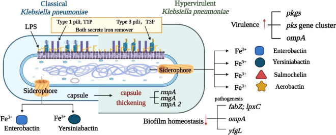
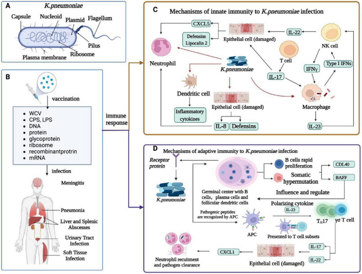
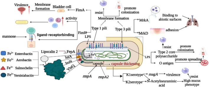
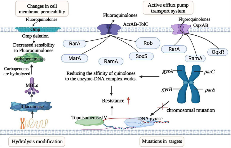
Klebsiella pneumoniae is a significant pathogenic bacterium responsible for a range of infections. The escalating prominence of K. pneumoniae in hospital-acquired infections is a deeply alarming trend that demands immediate attention and rigorous intervention. This article provides an up-to-date review of K. pneumoniae's virulence factors, pathogenesis, and the mechanism driving drug resistance. It also explores the potential for safe and effective vaccine developments, vital for preventing and controlling these diseases. Furthermore, we summarize the epidemiological characteristics of classical and hypervirulent K. pneumoniae infections, providing an overview of drug-resistance K. pneumoniae emergence, transmission, and prevalence.

Keywords: Klebsiella pneumoniae; drug resistance; epidemiology; pathogenesis; vaccine; virulence.


Conflict of interest statement

The authors declare that the research was conducted in the absence of any commercial or financial relationships that could be construed as a potential conflict of interest.

Figures

FIGURE 1
FIGURE 1
Comparison of virulence factors between classical and highly virulent K. pneumoniae. This figure illustrates a comparative analysis of virulence factors found in classical and highly virulent strains of K. pneumoniae. Four well-characterized virulence factors are highlighted: capsule, LPS, Pili (types 1 and 3), and siderophore. Notably, the siderophore is a feature shared with other pathogens such as Enterobacter, Salmonella, Yersinia, and Oxytocin. Understanding these distinctions in virulence factors is crucial in elucidating the varying pathogenicity and clinical outcomes associated with different K. pneumoniae strains.
FIGURE 2
FIGURE 2
Immune response of K. pneumoniae infection. (A) Cellular composition of K. pneumoniae. (B) K. pneumoniae infection in humans and related vaccines. K. pneumoniae infections include meningitis, pneumonia, liver and spleen abscesses, UTIs, and soft tissue infections. The vaccines against K. pneumoniae include whole-cell vaccine, capsular polysaccharide vaccine, LPS -related vaccine, protein vaccine, conjugate vaccine, ribosomal vaccine, reverse vaccine, DNA vaccine, and mRNA vaccine. (C) Innate immunity of K. pneumoniae infection. K. pneumoniae can induce innate immune system activation and interact with neutrophils, macrophages, dendritic cells, and epithelial cells. DC cells produce inflammatory cytokines, macrophages are a major cellular target of IFN γ, NK cells are a major source of IL-22, and IL-17 and IL-22 help in the clearance of Klebsiella by regulating the antimicrobial activity of the epithelium; Together they produce CXCL5 and Lipocalin 2, which recruits neutrophil, and epithelial cells produce defensins and IL-8, which recruits neutrophil. The alveolar macrophage produces inflammatory cytokines, IL-23, and type I IFN, and IL-23 activates NK cells to produce cytotoxic effects. (D) Adaptive immunity in humans caused by K. pneumoniae infection. Antigen-activated germinal center B cells undergo rapid proliferation and somatic hypermutation of their immunoglobulin variable genes, follicular dendritic cell in vivo secrete t-cell signals CD40L and b-cell activating factor (Baff) that affect and regulate b-cell survival and differentiation, and b-cell production of antibodies that kill bacteria. The APC recognizes the Antigen-presenting cell peptide and presents it to a subset of T cells in a local lymph node. Apcs release polarized cytokines (for example, IL-23) that can activate Th17 and γδ T cells. These T cell subsets produce IL-17 and IL-22 and stimulate airway epithelial cells expression to mediate chemokines (for example, CXCL1). Activated neutrophils engulf and kill bacteria, thereby enhancing K. pneumoniae clearance and local inflammation resolution.
FIGURE 3
FIGURE 3
Comparison of pathogenesis mechanism between classical and highly virulent K. pneumoniae. This figure provides an insightful comparison of the pathogenesis mechanisms observed in classical and highly virulent K. pneumoniae strains. Notable factors and mechanisms include the production of K1 and K2 capsular types, the presence of magA exclusively in K1 strains, and the role of sialic acid in enhancing anti-phagocytic activity. LPS is shown to promote bacterial dissemination in the bloodstream and colonization in internal organs through o-antigen resistance to serum killing and anti-phagocytosis by core polysaccharides. Furthermore, T1P and T3P contribute to strain virulence by forming biofilms, with T1P amplifying phagocytosis via FimH and T3P causing lung infections through various mechanisms. This figure also highlights iron metabolism as a critical factor, with siderophores (Enterocin, Salmonella, Yersinia, and Aerobacteriaceae) aiding in virulence by absorbing host iron. It’s noted that Enterobacter is inhibited by the host molecule lipocalin-2, whereas Yersinia, Salmonella, and aerobactinins are not, and specific transporters (FepA, IroN, YbtQ, and IutA) facilitate the transport of trivalent iron to bacterial cells in a siderophore-dependent manner.
FIGURE 4
FIGURE 4
Mechanisms of resistance in K. pneumoniae. This figure outlines the resistance mechanisms employed by K. pneumoniae, categorized into four main groups: (I) Permeability Reduction: Mutations or deletions, particularly in adventitial channels like porin Omp, lead to decreased permeability of the bacterial cell membrane. (II) Antibiotic Modification: K. pneumoniae exhibits resistance through the hydrolytic modification of antibiotics, notably β-lactamases that hydrolyze β-lactam antibiotics. (III) Target Structure Alterations: Resistance is also achieved by mutations and transformation of antibiotic target structures on the bacterial cell surface, reducing the affinity of antibiotic molecules. (IV) Efflux Pump Overexpression: Overexpression of efflux pumps, such as AcraB-TolC and OqxAB, contributes to resistance by reducing antibiotic accumulation within the bacterial cell. Transcriptional regulators like RarA, MarA, RamA, SoxS, Rob, and OqxAB are involved in regulating these efflux pumps. Understanding these resistance mechanisms is crucial for developing effective strategies to combat antibiotic-resistant K. pneumoniae infections.
FIGURE 5
FIGURE 5
Genetic mobile elements (TN4401 and NTEKPC) contained by blaKPC. In K. pneumoniae, the antibiotic resistance gene blaKPC is carried by genetic mobile elements, specifically TN4401 and NTEKPC. These elements are classified based on the presence of particular insertion sequences located upstream of blaKPC. NTEKPC is categorized into three groups: groups: NTEKPC-I, without insertion; NTEKPC-II, with insertion ΔblaTEM; NTEKPC-III, with insertion TN 5563/IS 6100. NTEKPC-I: This group lacks upstream insertions and can be further subdivided into AS-IA (e.g., prototype pKP048), -IB (e.g., pKPHS2), -IC (e.g., pKP13D), and -Id (e.g., pKPC-LKEC). This classification depends on the insertion sites of IS26 and the presence of ISkpn8. NTEKPC-II: These elements contain an insertion known as ΔblaTEM. NTEKPC-II further divides into -IIa (e.g., pFP10-1), -IIb (identified in strains M9884 and M9988), and -IIc (e.g., PPA-2). These subcategories are determined by variations in the length and deletions associated with △ blaTEM. Understanding these genetic mobile elements is crucial for unraveling the mechanisms of antibiotic resistance conferred by blaKPC in different K. pneumoniae strains. These insights are invaluable in the fight against antibiotic resistance in clinical contexts.
FIGURE 6
FIGURE 6
The transmission mechanism of resistance in K. pneumoniae. (A) The spread of resistance genes. qnr gene and aminoglycoside acetyltransferase variant gene [AAC(6′)-Ib-c] are commonly found in multiple resistance plasmids. Three quinolone resistance mediators are QNR protein, which can be divided into QnrA, QnrB, and QnrS, they protect against quinolone inhibition leading to resistance to the target enzyme encoding DNA helicase and topoisomerase IV; whereas a variant of the aminoglycoside-modified acetyltransferase AAC(6′)-Ib acetylates certain quinolones, reducing antibiotic activity leading to resistance, and then completes dissemination by plasmid transfer into host cells. (B) The type of plasmid backbone containing blaKPC. The plasmids with different types of blaKPC included IncF, IncI, IncA/C, IncN, IncX, IncR, IncP, IncU, IncW, IncL/M, and Cole plasmids.
FIGURE 7
FIGURE 7
Challenges and advances in multidrug-resistant Klebsiella pneumoniae vaccination. (A) Challenges in vaccine formulation. Fully leveraging the potential of bioinformatics and bioengineering, develop novel vaccine formulations and delivery systems, and utilize computer-aided design to maximize the response to the threat of multidrug-resistant Klebsiella pneumoniae to human health. (B) Preclinical trials: Using mouse, rabbit, and primate animal models to evaluate the efficacy of vaccines to prevent and treat diseases and identify potential side effects. (C) Clinical trials: After successful preclinical studies, the vaccines enter the clinical trial stage, and the safety and immunogenicity of the vaccines are comprehensively evaluated through efficacy evaluation system. (D) Epidemiology Research: Collect data on the effectiveness of vaccines in reducing or treating human infection rates or severity, and use this data to optimize traditional drugs by combining adjuvants and new targets.

References

    1. Ahn D., Prince A. (2020). Participation of the IL-10RB related cytokines, IL-22 and IFN-lambda in defense of the airway mucosal barrier. Front. Cell. Infect. Microbiol. 10:300. 10.3389/fcimb.2020.00300 - DOI - PMC - PubMed
    1. Alaubydi M., Al-Saadi D., Ameri L., Fadhil D. (2018). A novel method of preparation attenuated Klebsiella pneumoniae vaccine isolated from respiratory tract infections by using low power laser diodes. J. Mol. Microbiol. Biotechnol. 6 1441–1446.
    1. Amereh F., Arabestani M. R., Hosseini S. M., Shokoohizadeh L. (2023). Association of qnr genes and OqxAB Efflux pump in fluoroquinolone-resistant Klebsiella pneumoniae strains. Int. J. Microbiol. 2023:9199108. 10.1155/2023/9199108 - DOI - PMC - PubMed
    1. Arato V., Raso M. M., Gasperini G., Berlanda F., Scorza H., Micoli F. (2021). Prophylaxis and tareatment against Klebsiella pneumoniae: Current insights on this emerging anti-microbial resistant global threat. Int. Mol. J. Sci. 22:4042. 10.3390/ijms22084042 - DOI - PMC - PubMed
    1. Assoni L., Girardello R., Converso T. R., Darrieux M. (2021). Current stage in the development of Klebsiella pneumoniae Vaccines. Infect. Dis. Ther. 10 2157–2175. 10.1007/s40121-021-00533-4 - DOI - PMC - PubMed

LinkOut - more resources