. 2025 May 20:32:101881.
doi: 10.1016/j.mtbio.2025.101881. eCollection 2025 Jun.

Engineering 3D-BMSC exosome-based hydrogels that collaboratively regulate bone microenvironment and promote osteogenesis for enhanced cell-free bone regeneration

Affiliations

Engineering 3D-BMSC exosome-based hydrogels that collaboratively regulate bone microenvironment and promote osteogenesis for enhanced cell-free bone regeneration

Kai Xiang et al. Mater Today Bio. .

Abstract

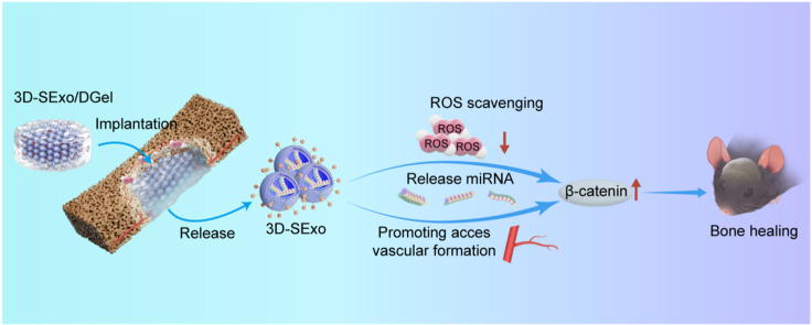
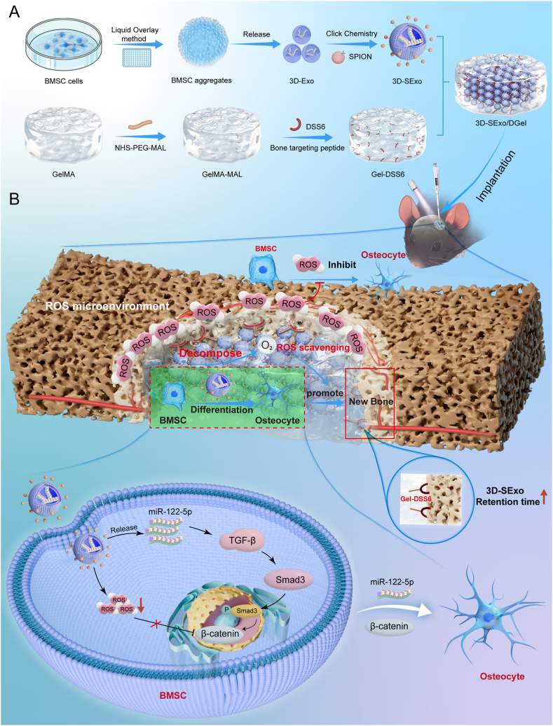
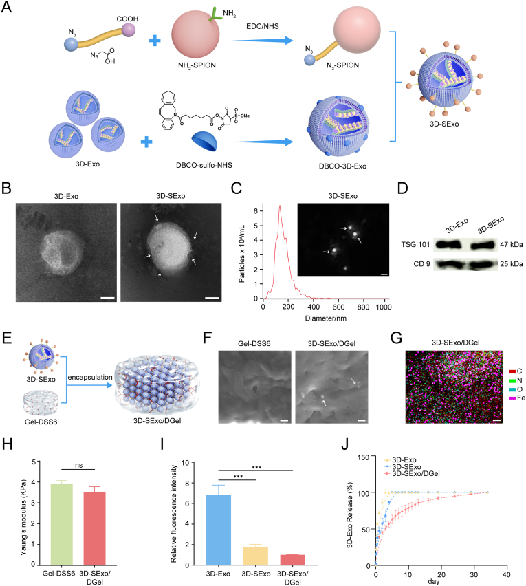
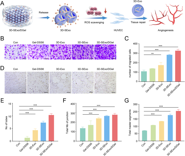
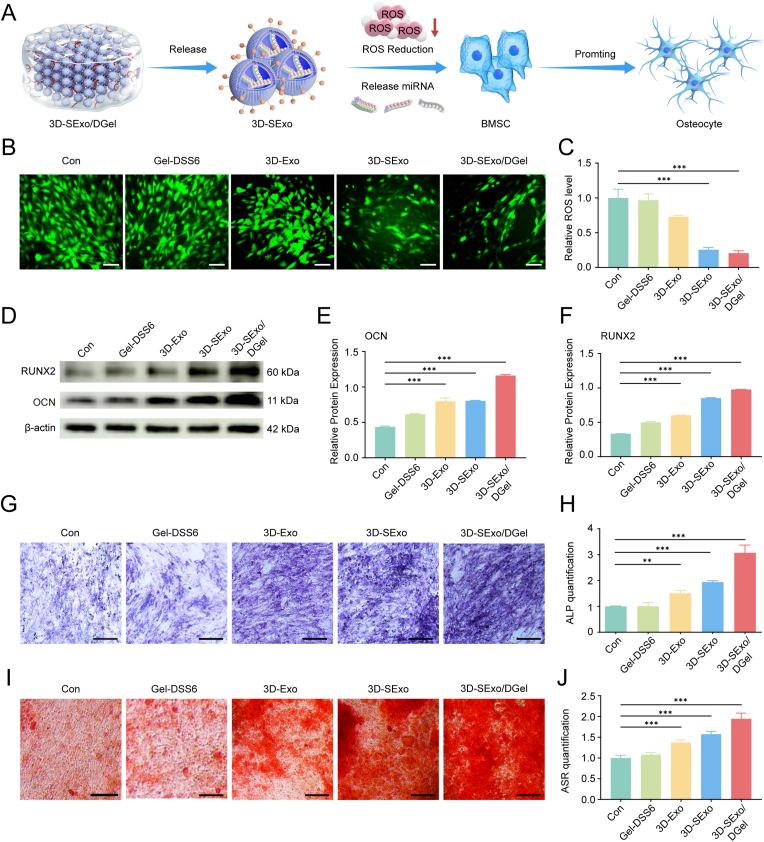
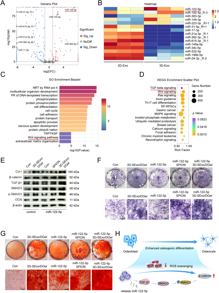
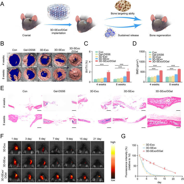
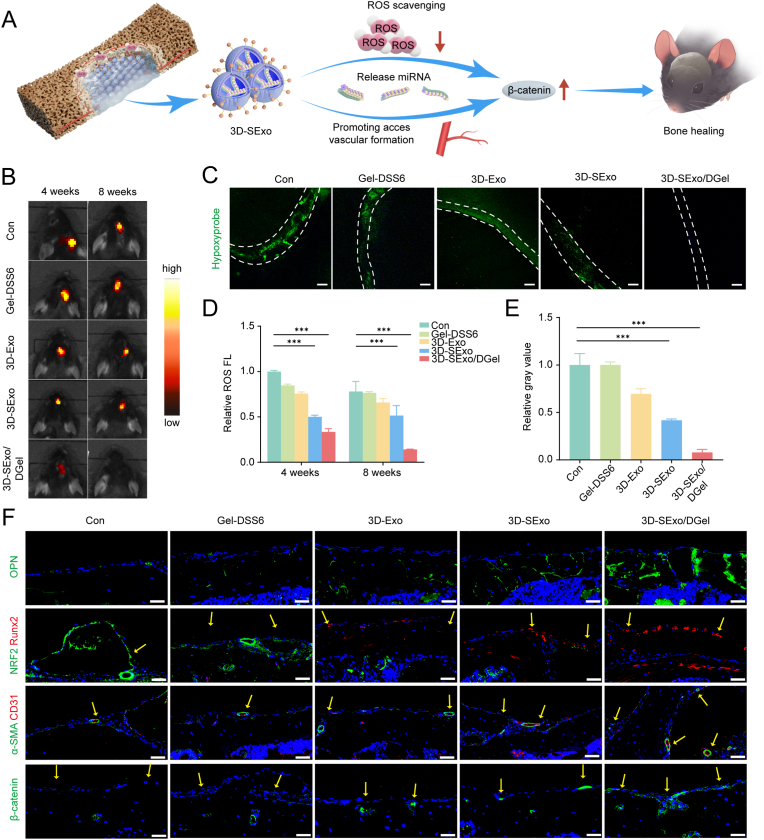
Large bone defects are a significant clinical challenge due to their frequent failure to heal spontaneously. Recently, BMSC-derived exosomes (Exo) based cell-free bone regeneration offer several distinct advantages over BMSCs themselves in the repair of damaged tissue, including enhanced repair ability and superior biocompatibility, which can be further augmented under 3D-cultured conditions. However, their therapeutic efficacy for bone regeneration is significantly constrained by hypoxic bone microenvironment and short retention time in bone defect region. Thus, judiciously regulating bone microenvironment and extending retention time are crucial for bone regeneration. Herein, we developed Superparamagnetic Iron Oxide Nanoparticles (SPION) -modified 3D-cultured Exo, termed as 3D-SExo, to enhance Reactive Oxygen Species (ROS) scavenging and promote bone regeneration in response to the needs of bone defect. After entrapment in bone-targeting peptide-modified GelMA (Gel-DSS6), the composite hydrogel (3D-SExo/DGel) was obtained, which can prolong the retention of exosomes, and thereby enhancing bone repair ability. In addition, miR-122-5p, detected via microRNA (miRNA) array from 3D-cultured Exo, were observed to promote osteogenesis by activating Wnt/β-catenin pathway, which was further verified by miRNA transfection. Through the in vitro and in vivo studies, 3D-SExo/DGel could decompose ROS to relive hypoxia and alleviate the inhibitory effect of ROS on β-catenin production, demonstrating significant clinical therapeutic potential to improve cell-free bone regeneration.

Keywords: Bone-targeted hydrogels; Collaboratively promote osteogenesis; Engineered 3D-BMSC Exosome; Enhanced cell‐free bone regeneration; Reactive oxygen species; Wnt/β-catenin pathway.


Conflict of interest statement

The authors declare that they have no known competing financial interests or personal relationships that could have appeared to influence the work reported in this paper.

Figures

Image 1
Graphical abstract
Fig. 1
Fig. 1
Schematic illustration of Engineering 3D-BMSC Exosome-based hydrogels with the capacity to collaboratively regulate bone microenvironment and promote osteogenesis for enhanced cell‐free bone regeneration thought activating Wnt/β-catenin pathway. (A) 3D-Exo were isolated from the serum-free supernatant of BMSC aggregates prepared by using a liquid overlay method. SPION were conjugated on the surface of 3D-Exo though click chemistry, then 3D-SExo were obtained. After encapsulated in bone targeting peptide (DSS6) conjugated GelMA (Gel-DSS6), the engineering 3D-BMSC Exosome-based hydrogels(3D-SExo/DGel) were constructed. (B) 3D-SExo/DGel can collaboratively regulate bone microenvironment and promote osteogenesis for enhanced cell‐free bone regeneration. In detail, after implanted into the bone defect region with 3D-SExo/DGel, 3D-SExo which were controlled-released from Gel-DSS6 could decompose ROS to relive hypoxia and alleviate the inhibitory effect of ROS on β-catenin production, and therefore enhanced the ability of 3D-Exo released microRNA (miR-122-5p) to promote osteogenesis by activating the Wnt/β-catenin pathway.
Fig. 2
Fig. 2
Synthesis and characterization of 3D-SExo/DGel hydrogel. (A) Schematic of the synthesis of 3D-SExo/DGel hydrogel. (B) Representative TEM images of 3D-Exo and 3D-SExo. Scale bar: 20 nm. (C) Nanoparticle tracking analysis of 3D-SExo. The insert image indicated the live stream views of 3D-SExo. Scale bar: 200 nm. (D)The expression of surface protein markers in 3D-Exo and 3D-SExo. (E) Schematic diagram of the construction of 3D-SExo/DGel. (F) Representative SEM images of Gel-DSS6 and 3D-SExo/DGel hydrogel. Scale bar: 200 nm. (G) Elemental mapping of 3D-SExo/DGel hydrogel. Scale bar: 100 μm. (H) Young's modulus of the hydrogel (n = 3). Data are shown as means ± SD (n = 3). The significant differences are determined by the unpaired t-test (double tail), NS: Not Statistically Significant, in comparison with the control group (Gel-DSS6). (I) The relative residual ROS fluorescence intensity of 3D-SExo/DGel after incubated with H2O2 for 12 h. The fluorescence intensity in the 3D-SExo/DGel group was normalized to 1. Data are shown as means ± SD (n = 3). The Residual ROS fluorescence intensity of 3D-SExo/DGel were set as 1. The significant differences are determined by the one-way ANOVA, ∗∗∗p < 0.005, in comparison with the control group (3D-SExo/DGel). (J) In vitro cumulative release of 3D-Exo from 3D-SExo/DGel hydrogel. Data are shown as means ± SD (n = 3).
Fig. 3
Fig. 3
3D-SExo/DGel hydrogel promotes angiogenesis in vitro. (A) Schematic diagram of 3D-SExo/DGel hydrogel promoting angiogenesis. (B) Representative images of HUVECs migration under hypoxia condition with different treatments. Scale bars: 50 μm. (C) Statistics of the number of migrated cells. Data are shown as means ± SD (n = 3). The significant differences are determined by the one-way ANOVA, ∗∗p < 0.01, ∗∗∗p < 0.005, in comparison with the control group. (D) Tube Formation of representative image networks in different groups of HUVECs. Scale bars:100 μm (E) Number of tubes of HUVEC under hypoxia condition with different treatments (n = 3). Data are shown as means ± SD (n = 3). The significant differences are determined by the one-way ANOVA, ∗∗∗p < 0.005, in comparison with the control group. (F) Total number of connections of HUVECs under hypoxia condition with different treatments. Data are shown as means ± SD (n = 3). The significant differences are determined by the one-way ANOVA, ∗∗∗p < 0.005, in comparison with the control group. (G) Total segments of HUVECs under hypoxia condition with different treatments. Data are shown as means ± SD (n = 3). The significant differences are determined by the one-way ANOVA, ∗∗∗p < 0.005, in comparison with the control group.
Fig. 4
Fig. 4
3D-SExo/DGel hydrogel promotes osteoblasts osteoblast differentiation under hypoxia condition in vitro. (A) Schematic diagram of 3D-SExo/DGel hydrogel in promoting osteoblast differentiation. (B) Representative images of ROS fluorescence staining after treatment of BMSC cells with different methods. Scale bars: 100 μm. (C) Quantitative analysis of ROS fluorescence staining. Data are shown as means ± SD (n = 3). The significant differences are determined by the one-way ANOVA, ∗∗∗p < 0.005, in comparison with the control group. (D) Western blot of OCN, RUNX2 and β-actin levels in different treated BMSC differentiated osteoblasts. (E–F) Quantitative analysis of relative OCN(E) and RUNX2 (F) in differentially differentiated osteoblasts from different treated BMSC (n = 3). Data are shown as means ± SD (n = 3). The significant differences are determined by the one-way ANOVA, ∗∗∗p < 0.005, in comparison with the control group. (G) Representative images of ALP staining in BMSC differentiated osteoblasts with different treatments. Scale bars: 100 μm. (H) Quantitative results of ALP staining in BMSC differentiated osteoblasts with different treatments. The expression level in the Con group was normalized to 1. Data are shown as means ± SD (n = 3). The significant differences are determined by the one-way ANOVA, ∗∗p < 0.01, ∗∗∗p < 0.005, in comparison with the control group. (I) Representative images of alizarin red staining in BMSC differentiated osteoblasts with different treatment. Scale bars: 100 μm. (J) Quantitative results of alizarin red staining in BMSC differentiated osteoblasts with different treatments. The expression level in the Con group was normalized to 1. Data are shown as means ± SD (n = 3). The significant differences are determined by the one-way ANOVA, ∗∗∗p < 0.005, in comparison with the control group. (For interpretation of the references to colour in this figure legend, the reader is referred to the Web version of this article.)
Fig. 5
Fig. 5
3D-SExo/DGel hydrogel promotes osteogenic differentiation in vitro via the Wnt/β-catenin pathway. (A) Volcano plot of the miRNAs in the 2D-Exo and 3D-Exo. (B) Heatmap of differentially expressed miRNAs between 2D-Exo and 3D-Exo. (C)Histogram of GO enrichment analysis. (D) Enriched pathways for target genes of miRNAs enriched within differential miRNAs in KEGG pathways. The size of the dot shows the number of differentially expressed genes in this pathway. The greater the rich factor value, the greater the degree of KEGG enrichment. (E) The expression of Col I, β-catenin, TGF-β1, SMAD3, p-SMAD3, OCN and β-actin levels in BMSC differentiated osteoblasts under hypoxia condition with or without miR-122-5p transfection after incubated with 3D-SExo/DGel hydrogel. (F) Representative images of ALP staining in BMSC differentiated osteoblasts under hypoxia condition with or without miR-122-5p transfection after incubated with 3D-SExo/DGel hydrogel. Scale bars: 100 μm. (G) Representative images of alizarin red staining in BMSC differentiated osteoblasts under hypoxia condition with or without miR-122-5p transfection after incubated with 3D-SExo/DGel hydrogel. Scale bars: 100 μm. (H) Schematic diagram of 3D-SExo/DGel promotes BMSC osteoblast differentiation under hypoxia condition through activation of Wnt/β-catenin pathway. (For interpretation of the references to colour in this figure legend, the reader is referred to the Web version of this article.)
Fig. 6
Fig. 6
3D-SExo/DGel hydrogel promotes osteogenesis in vivo. (A) Schematic representation of 3D-SExo/DGel hydrogel promote bone repair after implanted into the region of cranial bone defects in mice. (B) Representative 3D reconstructed micro-CT images of the cranial defect region in mice with different treatments. Red circles indicate the extent of the original bone defect. Scale bars: 500 μm. (C–D) Calculated bone volume fraction (C) and bone mineral density (D) in the cranial defect region from mice with different treatments (n = 3). Data are shown as means ± SD (n = 3). The significant differences are determined by the one-way ANOVA, ∗∗p < 0.01, ∗∗∗p < 0.005, in comparison with the control group. (E) Representative images of H&E staining in the cranial defect area of mice with different treatments. Scale bars: 300 μm for E. (F)In vivo imaging of DiR labeled 3D-SExo/DGel hydrogel after implanted into the region of cranial bone defects of mice. (G) Relative fluorescence intensity of cranial bone defect region of mice after implanted with DiR labeled 3D-SExo/DGel hydrogel. The fluorescence intensity of bone defect region implanted with DiR labeled formulation at day 1 were set as 1. Data are shown as means ± SD (n = 3). (For interpretation of the references to colour in this figure legend, the reader is referred to the Web version of this article.)
Fig. 7
Fig. 7
3D-SExo/DGel hydrogel regulate bone microenvironment to promote osteogenesis and angiogenesis in vivo. (A) Schematic illustration of ROS scavenging and angiogenesis in the anaerobic microenvironment of the cranial bone defect area, which promotes osteogenesis through activation of the Wnt/β-catenin pathway to promote the rise of catenin content and thus osteogenesis to promote osteogenic regeneration in vivo. (B) Representative images of the in vivo imaging system after injection of ROS Brite™ 700 at different time points. (C) Representative fluorescence images after injection of Hypoxyprobe in the cranial defect region at week eight. Scale bars: 500 μm. (D) Quantitative analysis of ROS Brite™ 700. The fluorescence intensity of the Con group at Week 4 was normalized to 1. Data are shown as means ± SD (n = 3). The significant differences are determined by the one-way ANOVA, ∗∗∗p < 0.005, in comparison with the control group. (E) Quantitative analysis of Hypoxyprobe staining. The fluorescence intensity of the Con group was normalized to 1. Data are shown as means ± SD (n = 3). The significant differences are determined by the one-way ANOVA, ∗∗∗p < 0.005, in comparison with the control group. (F) Representative images of immunofluorescence staining for OPN, NRF2, RUNX2, CD31, α-SMA, and β-catenin in the area of cranial defects in mice after different treatments at 8 weeks (yellow arrows point to the periosteum where osteoblasts are located). Scale bars: 50 μm. (For interpretation of the references to colour in this figure legend, the reader is referred to the Web version of this article.)

Similar articles

References

    1. Filippi M., Born G., Chaaban M., Scherberich A. Natural polymeric scaffolds in bone regeneration. Front. Bioeng. Biotechnol. 2020;8:474. - PMC - PubMed
    1. Perez J.R., Kouroupis D., Li D.J., Best T.M., Kaplan L., Correa D. Tissue engineering and cell-based therapies for fractures and bone defects. Front. Bioeng. Biotechnol. 2018;6:105. - PMC - PubMed
    1. Tarchala M., Harvey E.J., Barralet J. Biomaterial-stabilized soft tissue healing for healing of critical-sized bone defects: the masquelet technique. Adv. Healthc. Mater. 2016;5(6):630–640. - PubMed
    1. Quarto R., Mastrogiacomo M., Cancedda R., Kutepov S.M., Mukhachev V., Lavroukov A., Kon E., Marcacci M. Repair of large bone defects with the use of autologous bone marrow stromal cells, TN. Engl. J. Med. 2001;344(5):385–386. - PubMed
    1. Wang Y., Wang J., Gao R., Liu X., Feng Z., Zhang C., Huang P., Dong A., Kong D., Wang W. Biomimetic glycopeptide hydrogel coated PCL/nHA scaffold for enhanced cranial bone regeneration via macrophage M2 polarization-induced osteo-immunomodulation. Biomaterials. 2022;285 - PubMed

LinkOut - more resources