Correlation between early corneal edema and endothelial cell loss after phacoemulsification cataract surgery
- PMID: 40510852
- PMCID: PMC12158987
- DOI: 10.3389/fmed.2025.1562717
Correlation between early corneal edema and endothelial cell loss after phacoemulsification cataract surgery
Abstract
Purpose: This study aimed to investigate the correlation between early corneal edema and loss of corneal endothelial cells after phacoemulsification.
Methods: The corneal condition of each operated eye was observed using slit-lamp biomicroscopy at different time points, and the clinical score of corneal edema was determined. Central corneal thickness (CCT) and endothelial cell density (ECD) of each operative eye were measured by corneal endothelial microscopy at different time points.
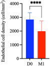
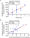
Results: There were 10 male (41.67%) and 14 female (58.33%) cataract patients with a mean age of aged 71.88 ± 10.14 years. The clinical score of corneal edema grade in cataract patients on the first postoperative day (1.79 ± 1.10) was significantly higher than that before surgery (0.0 ± 0.0) (p < 0.0001). Similarly, the CCT on first postoperative day (566.08 ± 32.73 μm) was significantly higher than that before surgery (530.71 ± 24.42 μm) (p < 0.0001). In addition, compared with the preoperative ECD (2,841 ± 502 cells/mm2), the ECD (1,993 ± 744 cells/mm2) 1 month after surgery (M1) decreased significantly (p < 0.0001), and the percentage of endothelial cell loss was 30.45 ± 20.23%. Moreover, both the corneal edema grade (Spearman's r = 0.7811, p < 0.0001) and CCT (Spearman's r = 0.7191, p < 0.0001) on the first day after surgery were significantly correlated with the loss of corneal endothelial cells 1 month postoperative day.
Conclusion: Early corneal edema after cataract surgery is closely associated with the loss of central corneal endothelial cells. Therefore, the grade of corneal edema and CCT on the first day after cataract surgery can be used as effective indices for predicting the effects of phacoemulsification on corneal endothelial cells.
Keywords: cataract; corneal edema; corneal endothelial cells; correlation; phacoemulsification.
Copyright © 2025 Chen, Li, Huang, Feng, Lu and Mu.
Conflict of interest statement
The authors declare that the research was conducted in the absence of any commercial or financial relationships that could be construed as a potential conflict of interest.
Figures


Similar articles
-
Femtosecond laser-assisted cataract surgery in Fuchs endothelial corneal dystrophy: Long-term outcomes.J Cataract Refract Surg. 2018 Jul;44(7):864-870. doi: 10.1016/j.jcrs.2018.05.007. Epub 2018 Jun 27. J Cataract Refract Surg. 2018. PMID: 29958766
-
[Effect of femtosecond laser-assisted phacoemulsification on corneal endothelium and prognosis of diabetic patients with cataract with different nuclear hardness].Zhonghua Yan Ke Za Zhi. 2024 Jun 11;60(6):511-517. doi: 10.3760/cma.j.cn112142-20231223-00305. Zhonghua Yan Ke Za Zhi. 2024. PMID: 38825950 Chinese.
-
Correlation Between Postoperative Central Corneal Thickness and Endothelial Damage After Cataract Surgery by Phacoemulsification.Cornea. 2018 May;37(5):587-590. doi: 10.1097/ICO.0000000000001502. Cornea. 2018. PMID: 29303887
-
Comparison of femtosecond laser-assisted cataract surgery and conventional phacoemulsification on corneal impact: A meta-analysis and systematic review.PLoS One. 2023 Apr 14;18(4):e0284181. doi: 10.1371/journal.pone.0284181. eCollection 2023. PLoS One. 2023. PMID: 37058458 Free PMC article.
-
Corneal Edema after Cataract Surgery.J Clin Med. 2023 Oct 25;12(21):6751. doi: 10.3390/jcm12216751. J Clin Med. 2023. PMID: 37959216 Free PMC article. Review.
References
LinkOut - more resources
Full Text Sources

