. 2025 Jun 5;15(11):e5330.
doi: 10.21769/BioProtoc.5330.

The Centriole Stability Assay: A Method to Investigate Mechanisms Involved in the Maintenance of the Centrosome Structure in Drosophila Cultured Cells

Affiliations

The Centriole Stability Assay: A Method to Investigate Mechanisms Involved in the Maintenance of the Centrosome Structure in Drosophila Cultured Cells

Mariana Lince-Faria et al. Bio Protoc. .

Abstract

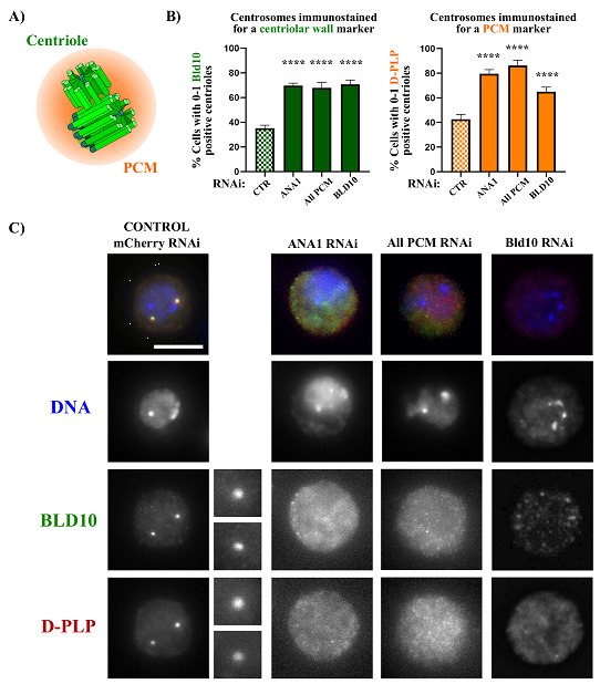
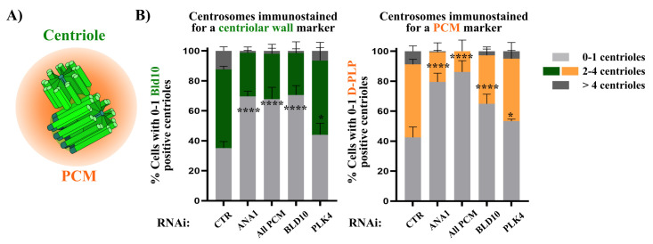
Centrosomes are vital eukaryotic organelles involved in regulating cell adhesion, polarity, mobility, and microtubule (MT) spindle assembly during mitosis. Composed of two centrioles surrounded by the pericentriolar material (PCM), centrosomes serve as the primary microtubule-organizing centers (MTOCs) in proliferating cells. The PCM is crucial for MT nucleation and centriole biogenesis. Centrosome numbers are tightly regulated, typically duplicating once per cell cycle, during the S phase. Deregulation of centrosome components can lead to severe diseases. While traditionally viewed as stable structures, centrosomes can be inactivated or disappear in differentiating cells, such as epithelial cells, muscle cells, neurons, and oocytes. Despite advances in understanding centrosome biogenesis and function, the mechanisms maintaining mature centrosomes or centrioles, as well as the pathways regulating their inactivation or elimination, remain less explored. Studying centrosome maintenance is challenging as it requires the uncoupling of centrosome biogenesis from maintenance. Tools for acute spatial-temporal manipulation are often unavailable, and manipulating multiple components in vivo is complex and time-consuming. This study presents a protocol that decouples centrosome biogenesis from maintenance, allowing the study of critical factors and pathways involved in the maintenance of the integrity of these important cellular structures. Key features • Drosophila cultured cells are resistant to centriole reduplication during S phase arrest, making them a suitable model for studying centrosome integrity without confounding effects from centriole biogenesis.

Keywords: Centrioles; Centrosomes; DMEL-2 cells; Drosophila melanogaster; Maintenance of centrosome integrity.


Conflict of interest statement

Competing interestsThe authors declare no competing interests.

Figures

Figure 1.
Figure 1.. Centriole stability assay showing that depletion of the centriolar wall protein ANA1 and the pericentriolar material (PCM) leads to centrosome loss.
(A) The centrosome is composed of two barrel-shaped microtubule-based centrioles surrounded by a multiprotein cloud called the PCM. (B) Centriolar numbers were assessed by considering the staining of the centriolar marker BLD10 (green bars) and the PCM marker D-PLP (orange bars). Plots showing the percentage of cells with abnormally low centriole numbers (0–1 centrioles) upon treatment with the different dsRNAs. We also included a condition in which BLD10 was depleted to validate the BLD10 antibody as a good centrosomal marker. Data are the mean ± SEM of three independent experiments (for each biological replicate in each experimental condition, n ≥ 100 cells). Statistical significance was determined by performing a bimodal regression test. The impact of the different RNAi treatments on the number of cells with 0–1 centrioles was estimated based on the number of cells that present a reduced number of centrioles with the different markers. Estimates indicate the log odds ratio that the indicated treatment increases the number of cells with 0–1 centrioles. *p < 0.05; **p < 0.01; ***p < 0.001; ****p < 0.0001; ns, not significant. Adapted from Pimenta-Marques et al. [18] (C) Representative images of centrosomes stained for the centriolar protein BLD10 (green) and the PCM protein D-PLP in control cells (mCherry RNAi) and cells with dsRNA transfection for the centriolar protein ANA1, the PCM (“All PCM;” ASL, CNN, D-PLP, and SPD2), and the centriolar protein BLD10. DNA (blue) enlargements of centrosomes present in each cell are shown. Scale bar, 5 μm.
Figure 2.
Figure 2.. Example of a 6-well plate layout for an assay where the function of individual proteins is tested for their role in centrosome stability.
In this setup, we recommend using 40 μg when single genes are silenced by the dsRNA. To ensure the same total amount of dsRNA in all conditions, for the positive control where the PCM is silenced (“All PCM”), we recommend the use of 10 μg of dsRNA to silence each gene, which is sufficient to induce defects in centrosome stability (Figure 1).
Figure 3.
Figure 3.. Example of a 6-well plate layout for an assay in which the function of individual proteins and their interaction are tested for a role in centrosome stability.
This assay includes the individual and simultaneous silencing of the hypothetical genes A and B. To compare phenotypes between single and simultaneous RNAi of genes A and B, the total amount of dsRNA silencing these genes (40 μg) must be equal in all wells (B1, B2, and B3). Therefore, when depleting A and B individually, we add 20 μg of mCherry dsRNA to have a total of 40 μg of dsRNA. The same rationale must be applicable to the negative and positive controls.
Figure 4.
Figure 4.. Quantification of centriole numbers in S phase arrested cells and depletion of different proteins of interest.
(A) Schematic representation of both centrioles (green) surrounded by the pericentriolar material (PCM) (orange). (B) Centriole numbers were quantified by independently counting the number of focused dots labeled with the centriole marker (BLD10) and the PCM marker (D-PLP). Centrioles may also be quantified based on the colocalization of both markers. Depletion of either ANA1 or the PCM (“All PCM;” ASL, CNN, D-PLP, and SPD2) leads to an increased percentage of cells with an abnormally low number of centrioles (0–1), as identified by each respective marker in comparison with the control (mCherry RNAi). Notably, RNAi for PLK4, a master regulator of centriole biogenesis, does not result in a substantial loss of centrioles. This contrasts sharply with the depletion or inhibition of PLK4 in cycling cells, which prevents centriole duplication and leads to centriole loss [27,33,34]. These results demonstrate the reliability of the centriole stability assay for investigating mechanisms that regulate the integrity of centrosomes. Note that a condition in which BLD10 is depleted was also included to validate the BLD10 antibody as a good centrosomal marker. Data are the mean ± SEM of three independent experiments (for each biological replicate in each experimental condition, n ≥ 100 cells). Statistical significance was determined by performing a bimodal regression test. The impact of the different RNAi treatments on the number of cells with 0–1 centrioles was estimated based on the number of cells that present a reduced number of centrioles with the different markers. Estimates indicate the log odds ratio that the indicated treatment increases the number of cells with 0–1 centrioles. *p < 0.05; ***p < 0.001; ****p < 0.0001; ns, not significant. Adapted from Pimenta-Marques et al. [18].

Similar articles

References

    1. Bettencourt-Dias M. and Glover D. M.(2007). Centrosome biogenesis and function: centrosomics brings new understanding. Nat Rev Mol Cell Biol. 8(6): 451 463 463. 10.1038/nrm2180 - DOI - PubMed
    1. Gönczy P. and Hatzopoulos G. N.(2019). Centriole assembly at a glance. J Cell Sci. 132(4): e228833. https://doi.org/ 10.1242/jcs.228833 - DOI - PubMed
    1. Pimenta-Marques A. and Bettencourt-Dias M.(2020). Pericentriolar material. Curr Biol. 30(12): R687–R689. 10.1016/j.cub .2020.04.064 - DOI - PubMed
    1. Vasquez-Limeta A. and Loncarek J.(2021). Human centrosome organization and function in interphase and mitosis. Semin Cell Dev Biol. 117: 30 41 41. 10.1016/j.semcdb .2021.03.020 - DOI - PMC - PubMed
    1. Balestra F. R., von Tobel L. and Gönczy P.(2015). Paternally contributed centrioles exhibit exceptional persistence in C. elegans embryos. Cell Res. 25(5): 642 644 644. 10.1038/cr.2015 .49 - DOI - PMC - PubMed

LinkOut - more resources