. 2025 Jun 13;14(6):516-526.
doi: 10.1302/2046-3758.146.BJR-2024-0287.R2.

Hederagenin promotes SIRT6 to attenuate epidural scar formation by aggravating PRMT1 deacetylation

Affiliations

Hederagenin promotes SIRT6 to attenuate epidural scar formation by aggravating PRMT1 deacetylation

Xiao-Chen Fan et al. Bone Joint Res. .

Abstract

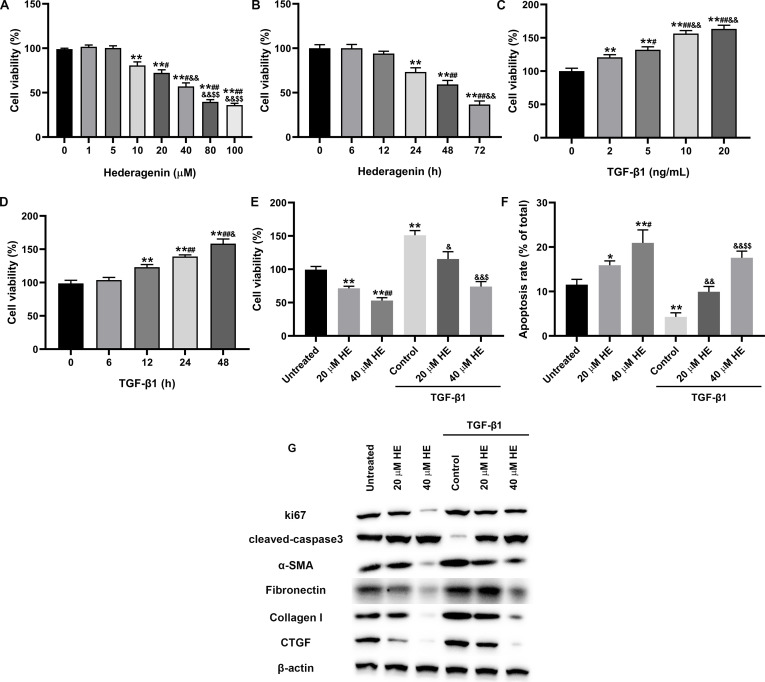
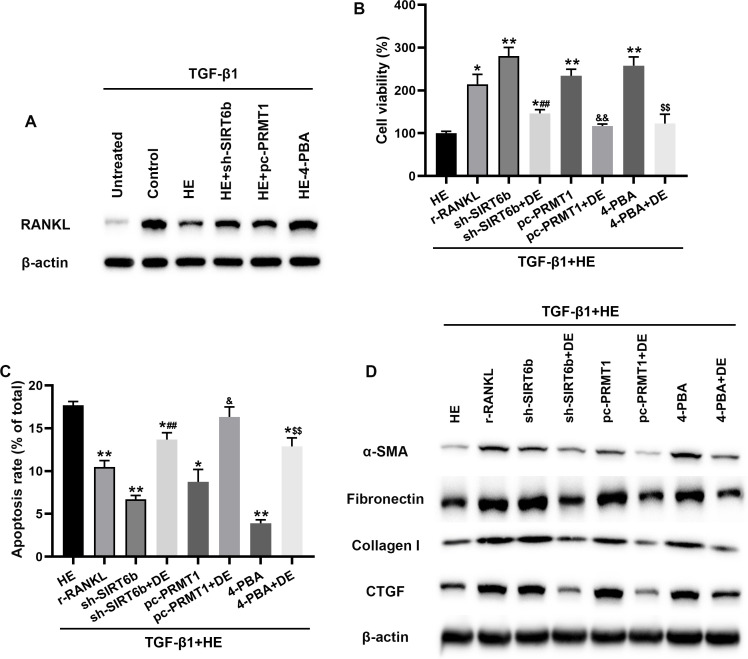
Aims: The formation of a postoperative epidural scar induced by epidural fibrosis is the main reason for recurrence of lumbar disc herniation after laminectomy. Hederagenin (HE) has been found to be widely present in various medicinal plants and has various pharmacological functions. This study aimed to investigate the effect and regulatory mechanism of HE on epidural scar formation.

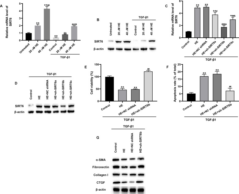
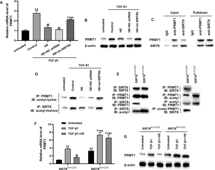
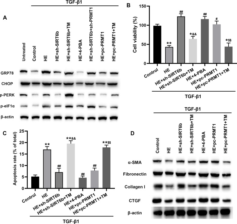
Methods: Transforming growth factor beta 1 (TGF-β1)-stimulated epidural scar fibroblasts were used as an in vitro cell model. Based on that, HE treatment was carried out along with sirtuin-6 (SIRT6) silence or protein arginine N-methyltransferase 1 (PRMT1) overexpression. The interaction between SIRT6 and PRMT1 was evaluated by pulldown and co-immunoprecipitation (CoIP) assays. Then, cell proliferation, apoptosis, and fibrosis were measured by Cell Counting Kit (CCK)-8, flow cytometry, and western blotting. Moreover, the effects of receptor activator of nuclear factor-κB ligand (RANKL) supplementation and endoplasmic reticulum (ER) stress were also evaluated by supplementing recombinant protein and specific inhibitor or activator.

Results: HE depressed cell proliferation and fibrosis, while inducing apoptosis of epidural fibroblasts. Meanwhile, HE promoted SIRT6 expression which suppressed PRMT1 acetylation and protein stability. Additionally, HE induced ER stress and upregulated RANKL in epidural fibroblasts via mediating SIRT6/PRMT1 axis.

Conclusion: Generally, the therapeutic role of HE treatment on epidural scar formation was exerted by regulating SIRT6/PRMT1 axis-mediated ER stress and RANKL pathway. This study provides evidence of a novel therapeutic measure for epidural scar formation.


Conflict of interest statement

J. Wang declares a grant from the National Natural Science Foundation of China for this study. X-C. Fan reports grants from the Key Research and Development Program of Shaanxi (grant numbers: 2022ZDLSF04-10, 2023-YBSF-617).

Figures

Fig. 1
Fig. 1
The effect of hederagenin (HE) on transforming growth factor beta 1 (TGF-β1)-stimulated fibrosis in epidural fibroblasts. a) to e) Cell Counting Kit (CCK)-8 method was performed to evaluate cell viability. f) Apoptosis rate of fibroblasts was measured using flow cytometry. g) The expressions of target proteins were evaluated by western blotting. *p < 0.05; **p < 0.01; #p < 0.05; ##p < 0.01; &p < 0.05; &&p < 0.01; $p < 0.05; $$p < 0.01. Data are presented as mean (standard error of the mean) derived from at least three experiments and analyzed by post-hoc test. α-SMA, alpha smooth muscle actin; CTGF, connective growth tissue factor.
Fig. 2
Fig. 2
The role of SIRT6 during epidural fibrosis progression in vitro. a) and c) The expression of SIRT6 messenger RNA (mRNA) was measured using real-time quantitative polymerase chain reaction (RT-qPCR). b), d), and g) Western blotting showing the protein levels of SIRT6 and fibrotic proteins. e) Cell Counting Kit-8 assay was used to evaluate cell viability. f) Cell apoptosis was determined using a Cell Death Annexin V/PI kit (BD Biosciences, USA). The results obtained from triple experiments are analyzed by post-hoc test and shown as mean (standard error of the mean). **p < 0.01; #p < 0.05; ##p < 0.01; &p < 0.05; &&p < 0.01; $$p < 0.01. α-SMA, alpha smooth muscle actin; CTGF, connective tissue growth factor; HE, hederagenin; NC shRNA, negative control short hairpin RNA; TGF-β1, transforming growth factor beta 1.
Fig. 3
Fig. 3
The interaction and association between SIRT6 and PRMT1. a), b), f), and g) The expressions of PRMT1 at messenger RNA (mRNA) and protein levels were measured using real-time quantitative polymerase chain reaction and western blotting. c) Pulldown assay was used to evaluate the interaction between SIRT6 and PRMT1. d) and e) Co-immunoprecipitation (Co-IP) determined the interactions between wild-type and mutant type of SIRT6 with PRMT1, acetyl-PRMT1, acetyl-lysine, and acetyl-histone. Experiments were conducted at least three times. The final data were analyzed by post-hoc test and shown as mean (standard error of the mean). **p < 0.01; ##p < 0.01; &&p < 0.01. NC shRNA, negative control short hairpin RNA; TGF-β1, transforming growth factor beta 1.
Fig. 4
Fig. 4
Endoplasmic reticulum (ER) stress was involved in hederagenin (HE)-induced epidural fibrosis therapy. a) and d) The activation of ER stress and expressions of fibrotic proteins were measured using western blotting assay. b) Cell Counting Kit-8 assay was used to evaluate the cell viability. c) Cell apoptosis was determined by flow cytometry. The statistical results were based on at least three experimental repeats analyzed by post-hoc test and are presented as mean (standard error of the mean). *p < 0.05; **p < 0.01; #p < 0.05; ##p < 0.01; &&p < 0.01; $$p < 0.01. 4-PBA, 4-phenylbutyrate; α-SMA, alpha smooth muscle actin; CHOP, C/EBP homologous protein; CTGF, connective growth tissue factor; PRMT1, protein arginine N-methyltransferase 1; SIRT6, sirtuin-6; TGF-β1, transforming growth factor beta 1; TM, tunicamycin.
Fig. 5
Fig. 5
Receptor activator of nuclear factor-κB ligand (RANKL) plays an important role in HE-induced epidural fibrosis therapy. a) and d) The expressions of RANKL and fibrotic proteins were measured using western blotting. b) Cell Counting Kit-8 method evaluated the viability of fibroblast cells. c) Flow cytometry was used to determine the cell apoptosis rate. Data were based on three experimental repeats analyzed by post-hoc test and are shown as mean (standard error of the mean). *p < 0.05; **p < 0.01; ##p < 0.01; &p < 0.05; &&p < 0.01; $$p < 0.01. 4-PBA, 4-phenylbutyrate; α-SMA, alpha smooth muscle actin; CTGF, connective growth tissue factor; DE, denosumab; PRMT1, protein arginine N-methyltransferase 1; SIRT6, sirtuin-6; TGF-β1, transforming growth factor beta 1.

Similar articles

References

    1. Williams MG, Wafai AM, Podmore MD. Functional outcomes of laminectomy and laminotomy for the surgical management lumbar spine stenosis. J Spine Surg. 2017;3(4):580–586. doi: 10.21037/jss.2017.10.08. - DOI - PMC - PubMed
    1. Sun Y, Ge Y, Fu Y, et al. Mitomycin C induces fibroblasts apoptosis and reduces epidural fibrosis by regulating miR-200b and its targeting of RhoE. Eur J Pharmacol. 2015;765:198–208. doi: 10.1016/j.ejphar.2015.08.002. - DOI - PubMed
    1. Dai J, Li X, Yan L, et al. The effect of suramin on inhibiting fibroblast proliferation and preventing epidural fibrosis after laminectomy in rats. J Orthop Surg Res. 2016;11(1):108. doi: 10.1186/s13018-016-0443-5. - DOI - PMC - PubMed
    1. Tian F, Dou C, Qi S, et al. Preventive effect of dexamethasone gelatin sponge on the lumbosacral epidural adhesion. Int J Clin Exp Med. 2015;8(4):5478–5484. - PMC - PubMed
    1. Wang S, Li X, Yan L, et al. Tamoxifen inhibits fibroblast proliferation and prevents epidural fibrosis by regulating the AKT pathway in rats. Biochem Biophys Res Commun. 2018;497(4):937–942. doi: 10.1016/j.bbrc.2018.01.032. - DOI - PubMed