. 2025 Jul 24;207(7):e0005625.
doi: 10.1128/jb.00056-25. Epub 2025 Jun 13.

The cell wall hydrolase MltG is essential to maintain cell wall homeostasis of Enterococcus faecalis

Affiliations

The cell wall hydrolase MltG is essential to maintain cell wall homeostasis of Enterococcus faecalis

Alexis A U Knotek et al. J Bacteriol. .

Abstract

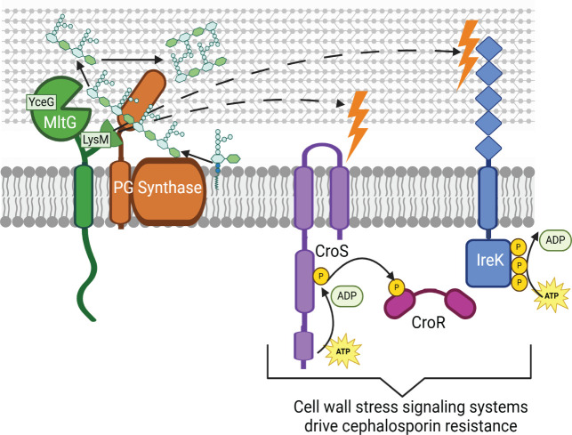
Infections caused by enterococci are increasingly prevalent and difficult to treat due to multidrug resistance. Enterococcus faecalis exhibits intrinsic resistance toward cephalosporins, which inhibit the final step of peptidoglycan (PG) synthesis. Intrinsic resistance requires multiple factors in the PG synthesis pathway and at least two cell-wall-stress signal transduction systems; however, the complete molecular mechanism of enterococcal cephalosporin resistance remains to be elucidated. MltG, a predicted PG hydrolase, is thought to process nascent strands of PG, suggesting that MltG might play an important role in enterococcal cell wall homeostasis and potentially cephalosporin resistance. Here, we demonstrate that enterococcal MltG cleaves nascent PG. An E. faecalis mutant lacking MltG exhibits several related phenotypes in the absence of exogenous stress: a marked growth defect, a loss of cell wall integrity, a reduction in PG synthesis, and activation of two cell-wall-stress signal transduction systems that drive elevated cephalosporin resistance. Together, these results are consistent with the model that MltG promotes proper cell wall homeostasis in E. faecalis, and further reveal that the enzymatic activity of MltG is not necessary for it to perform this function-instead, the LysM (putative PG-binding) domain of MltG plays the critical role. Nevertheless, the enzymatic activity of MltG does impact cephalosporin resistance, because a catalytically inactive MltG variant leads to elevated resistance. Collectively, our findings represent the first description of MltG function in E. faecalis and point to at least two distinct roles for MltG in PG homeostasis and cephalosporin resistance.

Importance: Enterococcus faecalis is an opportunistic pathogen that colonizes the human gut microbiome. Infections caused by E. faecalis are increasingly prevalent and difficult to treat due to the multidrug resistance exhibited toward common clinical antibiotics. A thorough understanding of the mechanisms used by E. faecalis to maintain cell wall homeostasis will serve as a foundation for future development of new therapeutics that disable enterococcal resistance to cell-wall-active antibiotics and may reveal new vulnerabilities that could be exploited by novel antimicrobials. Here, we demonstrate that the MltG peptidoglycan hydrolase is essential for enterococcal cell wall homeostasis, but that the enzymatic activity of MltG is not required for this role. Instead, the enzymatic activity of MltG impacts intrinsic resistance toward cephalosporins.

Keywords: Enterococcus; MltG; cell wall stress; cephalosporin resistance; peptidoglycan.


Conflict of interest statement

The authors declare no conflict of interest.

Figures

Fig 1
Fig 1
Enterococcal MltG cleaves nascent PG products in vitro. (A) Schematic of the catalytic activity assay used to visualize PG cleavage by MltG. Briefly, purified Pbp1A was incubated with Lipid II, followed by the addition of either wild-type (WT) MltG or MltG E342Q. PG products were then labeled with Biotinylated D-Lysine and run on 4%–20% gradient SDS-PAGE gel, and PG was detected with IR-streptavidin. (B) SDS-PAGE analysis of reactions with the indicated composition.
Fig 2
Fig 2
MltG deletion results in a growth defect. Bacterial growth (OD600) was monitored over the course of 24 hours. Data represent the mean ± standard deviation of three biological replicates (two replicates for WT/vector). Error bars are too small to be seen in some cases. Strains were (A) WT/vector = OG1(pJRG9); ΔmltG/WT mltG = JL650(pAAU12); ΔmltG/vector = JL650(pJRG9); (B) ΔmltG/WT mltG = JL650(pAAU12); ΔmltG/mltG E342Q = JL650(pAAK15); ΔmltG/vector = JL650(pJRG9); (C) ΔmltG/WT mltG = JL650(pAAU12); ΔmltG/WT mltG-his = JL650(pAAK33); ΔmltG/mltGEfm-his = JL650(pAAK76); ΔmltG/mltGEc-his = JL650(pAAK75); ΔmltG/vector = JL650(pJRG9); and (D) ΔmltG/WT mltG = JL650(pAAU12); ΔmltG/mltG ΔYceG = JL650(pAAK65); ΔmltG/mltG ΔLysM = JL650(pAAK67); ΔmltG/vector = JL650(pJRG9).
Fig 3
Fig 3
MltG deletion leads to a loss of cell wall integrity. Strains constitutively producing β-galactosidase were grown for 24 hours in MHB in the presence of CPRG (β-galactosidase substrate). CPRG cleavage (OD570) normalized to bacterial growth (OD630) is presented. n = 3 and error bars represent ±s.d. Strains were WT/vector = OG1(pJRG9); ΔmltG/vector = JL650(pJRG9); ΔmltG/WT mltG = JL650(pAAU12); ΔmltG/mltG E342Q = JL650(pAAK15).
Fig 4
Fig 4
MltG deletion triggers cell wall stress signaling. For each pairwise comparison, whole-cell lysates were subjected to phos-tag SDS-PAGE and then immunoblotting for IreK or CroR as indicated. The upper band(s) represent phosphorylated proteoform(s). Intensities from the phosphorylated and unphosphorylated signals were used to calculate % phosphorylation of IreK or CroR, presented as the average % phosphorylation in bar graphs. (A) Comparison of WT vs ΔmltG reveals that MltG deletion triggers phosphorylation of IreK and CroR. Comparison of (B) WT/vector vs ΔmltG/WT mltG or (C) ΔmltG expressing either WT mltG or ΔmltG /mltG E342Q reveals complementation of the ΔmltG mutation. n = 3 and error bars represent ±s.d. *P < .05; ns = not significant. Student’s t-test (heteroscedastic, two-tailed). Strains were WT = OG1; ΔmltG = JL650; ΔireK = JL206; ΔcroR = SB23; WT/vector = OG1(pJRG9); ΔmltG/WT mltG = JL650(pAAU12); ΔmltG/mltG E342Q = JL650(pAAK15).
Fig 5
Fig 5
MltG deletion increases PBP expression. Strains grown to exponential phase were exposed to Bocillin-FL, which acylates active PBPs. (A) Labeled cells were then lysed and subjected to SDS-PAGE. The image is representative of three biological replicates. (B) The intensity of Bocillin-FL labeling for each PBP was normalized to total protein staining (GelCode Blue). Graphs represent the average of three biological replicates. (C) Cell lysates were subjected to SDS-PAGE supplemented with TCE for total protein detection and immunoblot with antiserum for PbpA or Pbp4. Graphs represent the average of three biological replicates. Error bars represent ±s.d. *P< .05; Student’s t-test (heteroschidastic, two-tailed). Strains were WT/vector = OG1(pJRG9); ΔmltG/vector = JL650(pJRG9); ΔpbpA/vector = JL632(pJRG9); Δpbp4/vector = JL339(pJRG9).
Fig 6
Fig 6
MltG deletion reduces the rate of PG synthesis. [14C] GlcNAc incorporation into PG during pulse-labeling, normalized to cell density. Label incorporation rates presented in the table were determined from data points between 10 and 25 minutes. Data represent ±s.d. for two biological replicates. Strains were WT/vector = OG1(pJRG9); ΔmltG/vector = JL650(pJRG9); ΔmltG/WT mltG = JL650(pAAU12); ΔmltG/mltG E342Q = JL650(pAAK15).
Fig 7
Fig 7
Summary model. Our data support a model in which E. faecalis MltG cleaves nascent strands of PG through the action of its C-terminal YceG domain, presumably to enable proper incorporation of nascent PG strands into mature PG. In addition, the membrane-proximal LysM domain of MltG appears essential for the ability of MltG to support normal growth and cell wall integrity, because loss of the LysM domain (but not the catalytic YceG domain) results in activation of the CroRS and IreK cell wall stress signaling systems to drive hyper-resistance toward cephalosporins. Created in BioRender. Uitenbroek, A. (2025) https://BioRender.com/2l14ekc

References

    1. Hayashi H, Takahashi R, Nishi T, Sakamoto M, Benno Y. 2005. Molecular analysis of jejunal, ileal, caecal and recto-sigmoidal human colonic microbiota using 16S rRNA gene libraries and terminal restriction fragment length polymorphism. J Med Microbiol 54:1093–1101. doi: 10.1099/jmm.0.45935-0 - DOI - PubMed
    1. Kristich C, Rice L, Arias C. 2014. Enterococcal infection-treatment and antibiotic resistance. In Gilmore M, Clewell D, Ike Y, Shankar N (ed), Enterococci: from commensals to leading causes of drug resistant infection. Massachusetts Eye and Ear Infirmary. - PubMed
    1. Hollenbeck BL, Rice LB. 2012. Intrinsic and acquired resistance mechanisms in enterococcus. Virulence 3:421–433. doi: 10.4161/viru.21282 - DOI - PMC - PubMed
    1. Miller WR, Munita JM, Arias CA. 2014. Mechanisms of antibiotic resistance in enterococci. Expert Rev Anti Infect Ther 12:1221–1236. doi: 10.1586/14787210.2014.956092 - DOI - PMC - PubMed
    1. Brandl K, Plitas G, Mihu CN, Ubeda C, Jia T, Fleisher M, Schnabl B, DeMatteo RP, Pamer EG. 2008. Vancomycin-resistant enterococci exploit antibiotic-induced innate immune deficits. Nature 455:804–807. doi: 10.1038/nature07250 - DOI - PMC - PubMed

MeSH terms

LinkOut - more resources