. 2025 Jul 9;16(7):e0107625.
doi: 10.1128/mbio.01076-25. Epub 2025 Jun 13.

Structure and stabilization of the antigenic glycoprotein building blocks of the New World mammarenavirus spike complex

Affiliations

Structure and stabilization of the antigenic glycoprotein building blocks of the New World mammarenavirus spike complex

Guido C Paesen et al. mBio. .

Abstract

The spillover of New World (NW) arenaviruses from rodent reservoirs into human populations poses a continued risk to human health. NW arenaviruses present a glycoprotein (GP) complex on the envelope surface of the virion, which orchestrates host cell entry and is a key target of the immune response arising from infection and immunization. Each protomer of the trimeric GP is composed of a stable signal peptide, a GP1 attachment glycoprotein, and a GP2 fusion glycoprotein. To glean insights into the architecture of this key therapeutic target, we determined the crystal structures of NW GP1-GP2 heterodimeric complexes from Junín virus and Machupo virus. Due to the metastability of the interaction between GP1 and GP2, structural elucidation required the introduction of a disulfide bond at the GP1-GP2 complex interface, but no other stabilizing modifications were required. While the overall assembly of NW GP1-GP2 is conserved with that presented by Old World (OW) arenaviruses, including Lassa virus and lymphocytic choriomeningitis virus, NW GP1-GP2 complexes are structurally distinct. Indeed, we note that when compared to the OW GP1-GP2 complex, the globular portion of NW GP1 undergoes limited structural alterations upon detachment from its cognate GP2. We further demonstrate that our engineered GP1-GP2 heterodimers are antigenically relevant and recognized by neutralizing antibodies. These data provide insights into the distinct assemblies presented by NW and OW arenaviruses, as well as provide molecular-level blueprints that may guide vaccine development.IMPORTANCEAlthough the emergence of New World (NW) hemorrhagic fever mammarenaviruses poses an unceasing threat to human health, there is a paucity of reagents capable of protecting against the transmission of these pathogens from their natural rodent reservoirs. This is, in part, attributed to our limited understanding of the structure and function of the NW glycoprotein spike complex presented on the NW arenavirus surface. Here, we provide a detailed molecular-level description of how the two major components of this key therapeutic target assemble to form a key building block of the NW arenaviral spike complex. The insights gleaned from this work provide a framework for guiding the structure-based development of NW arenaviral vaccines.

Keywords: arenavirus; glycoprotein; rational immunogen design; structure; virus-host interactions.


Conflict of interest statement

The authors declare no conflict of interest.

Figures
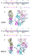
Fig 1
Fig 1
Structures of JUNV GP1−GP2 (top) and MACV GP1−GP2 (bottom). (A) Linear representation of the translation product of the JUNV spike gene, showing the SSP in gray, GP1 in violet, and GP2 in pale blue. The black triangles indicate the SPase and SKI-1 cleavage sites, and the pins represent N-linked glycosylation sequons. Pins are colored black if the glycan was observed to be ordered and occupied, gray if not. The strap domain is indicated, as are the fusion peptides (ƒ), the heptad repeat regions (HR1 and HR2 [30]), the T-loop, the membrane-proximal external region (MPER), the transmembrane domain (TM), and the intra-virion (cytosolic) domain (IV). Disulfide bonds are indicated by yellow brackets. (B) Linear representation of the JUNV GP188-329GP2e construct. The signal peptide and Twin-Strep tag are not shown. The added disulfide bond is indicated by the yellow bracket, and the introduced furin site by the black triangle. A proline (E321P) was also included, as incorporated in the study of LASV GP structure (13). The TM and IV regions were not included in the constructs. (C) Crystal structure of the JUNV GP1−GP2e protein in complex with Fab fragments of neutralizing monoclonal antibody (mAb) JUN1. JUNV GP1−GP2 is shown in cartoon representation, with GP in magenta and GP2 in blue. The Fab is shown as ribbons, with the heavy chain in gold and the light chain in black. Glycans are shown as sticks and colored according to the location of their sequons. Disulfide bonds are shown as yellow sticks. The inset shows a close-up of the GP1−GP2 interface, with the GP1 chain in pink, except for the N-terminal strap (magenta) and the C-terminal α4-helix (red). The GP2 chain is colored blue, except for the T-loop region (cyan). The added cysteines forming the inter-chain disulfide bond are labeled. (D and E) Linear representation of the translation product of the MACV spike gene and of the MACV GP188-340GP2e construct, respectively, using the same color scheme and symbols as in panels A and B, but with E→P denoting an E340P mutation. The mutation of Glu258 into a serine N-terminal of the furin site in the MACV construct introduced an extra NXS glycosylation sequon (pink pin) to the “SKI-loop.” This loop is not seen in the crystal structure, so any bound glycans are not visible. (F) Crystal structure of the MACV GP1−GP2e protein (cartoon) in complex with Fab fragments of neutralizing mAb MAC1 (ribbon). Colors and labeling are as in panel C.
Fig 2
Fig 2
Binding of JUNV- and MACV-specific mAbs to JUNV GP188-329GP2 and MACV GP188-340GP2 as measured by enzyme-linked immunosorbent assay (ELISA). mAbs used in this study include JUNV-specific neutralizing mAbs, JUN1 (39), JUN5 (39), JUN7 (39), OD01 (41, 45, 46), and the MACV-specific mAb, MAC1 (39). Measurements were performed in duplicate.
Fig 3
Fig 3
Structure-based classification of NW and OW arenaviral GP1 glycoproteins illustrates the similarity of NW GP1 glycoproteins in GP2-attached and -detached states. A pairwise distance matrix was calculated with Structural Homology Program (47–49). Pairwise evolutionary distance matrices were used to generate unrooted phylogenetic trees in PHYLIP (50). JUNV GP1, in the GP2-attached state, is shown in cartoon representation and colored as a rainbow from the N-terminus (blue) to C-terminus (red). Cartoon tube coloring from blue to orange, with increasing tube thickness, reflects increased structural distance from JUNV GP1 in the GP2-attached state upon overlay. Non-equivalent residues are colored red with exaggerated thickness. Structures used in the analysis are as follows: JUNV GP1 detached (PDB ID 5NUZ), JUNV GP1 attached (PDB ID 9GHJ), MACV GP1 attached (PDB ID 9GHI), MACV GP1 detached (PDB ID 2WFO), Whitewater Arroyo virus (WWAV) GP1 detached (PDB ID 6HJ4), Lujo virus (LUJV) GP1 detached (PDB ID 6GH8), LCMV GP1 attached (PDB ID 5INE), LASV GP1 attached (PDB ID 5VK2), Loei river virus (LORV) GP1 detached (PDB ID 6HJC), Morogoro virus (MORV) GP1 detached (PDB ID 5NFF), LASV GP1 (detached 4ZJF). A single asterisk indicates structures determined in this study. A double asterisk indicates LCMV GP1, as determined within a single GP2-attached heterodimer (PDB ID 5INE). A triple asterisk indicates LCMV GP1, as it occurs in a trimer of GP2-attached heterodimers. The LASV GP1 attached structure was determined as part of a trimer of GP1−GP2 heterodimers. All other structures were determined as either GP1−GP2 heterodimers (attached) or as free GP1 (detached).
Fig 4
Fig 4
The N-terminal GP1 strap. (A) Overview of interactions between the GP1 strap and GP2. Given that the straps of JUNV and MACV are similar, the strap of JUNV was chosen for comparison with LASV. The GP1 strap is shown as a magenta cartoon, with side chains shown as sticks. GP2 is shown in surface representation, with basic and acidic patches colored blue and red, respectively. Residues occupying the long groove on the GP2 surface are labeled with bold letter types for Phe75 (in black), Phe70 (purple), Phe62 (orange), and Ile64 (blue). Labels for the helices and strands at the GP1−GP2 interface are highlighted in green. (B) Binding of Phe75 of the strap to a conserved, hydrophobic pocket in GP2. Strap residues are shown in magenta, GP2 residues in light blue or, if they belong to the T-loop, in cyan. Hydrogen bonds are shown as orange lines, hydrophobic interactions as violet dashed lines. GP2 residues forming the pocket are conserved in NW and OW viruses, but Phe75 of the strap is frequently replaced by a leucine, isoleucine, or valine (see Fig. S7). (C) Comparison of the interactions at the N-terminal tip of the strap between the JUNV (a, b) and LASV (c, d) structures. In JUNV, Phe70 occupies a rather shallow, hydrophobic pocket, which consists of mostly aromatic GP2 residues (a). GP1 residues Phe62 and Ile64 join the pocket. Trp395 (labeled in red) plays a central role in the interaction, forming connections with Phe70 and Ile64, and π–stacking interactions with Phe62 (indicated by the green dashed lines and spheres denoting the aromatic ring centers). Trp395 and Ile388 link the C-terminal helix of the ectodomain (α9; b) to the N-terminal β-strands of the strap (β1, β2; the N-terminus is denoted by “N”). Interactions in LASV (PDB 7PUY) at the level of Ile68 (equivalent to Phe70 of JUNV) are markedly different (c, d, and Fig. S7). Ile403 (in red), which is strictly conserved in OW viruses, takes the place of the much larger Trp395 (fully conserved in NW viruses) on the C-terminal helix (α11; d). The smaller Ile403 partners with the aromatic Tyr62, which is also conserved in OW viruses and replaces Ile64, the smaller, NW-specific partner of Trp395. Phe62 of JUNV, another residue that interacts with Trp395 and which is also conserved as a hydrophobic residue in NW viruses, is replaced by Ser60 in LASV, which is not conserved in OW viruses and does not contribute to the strap-GP2 interaction, as observed in PDB ID 7PUY (shown as lines). Similarly, Trp386 (Trp378 in JUNV), Phe399 (Phe391), and Leu372 (Val364) do not bind the N-terminus of the strap in LASV.
Fig 5
Fig 5
Interactions at the C-terminal region of the strap. (A) Interactions between the C-terminal part of the strap domain in JUNV and the α6 helix of GP2, which in turn interacts with the structurally ordered portion of the N-terminal fusion peptide segment (N-FPS). The strap domain is colored magenta, GP2 domains are colored blue. The side chains of selected residues and main-chain atoms that form inter-chain hydrogen bonds are shown as sticks. Strap residues are labeled in bold. Hydrogen bonds are shown as solid orange lines, hydrophobic interactions as violet dotted lines, and π–stacking interactions as green dashed lines with spheres denoting the aromatic ring centers. (B) Direct interactions between the C-terminal part of the strap in JUNV and the internal fusion peptide segment (i-FPS). (C) Interactions between the C-terminal part of the strap and the i-FPS in MACV. The purple arrow indicates the orientation of α-helix 1, which contrasts with that observed in JUNV GP1. Instead of Asn83 interacting with i-FPS, as observed in JUNV GP1−GP2, Asn83 in MACV GP1−GP2 is distal from the fusion peptide and glycosylated. N-linked glycosylation is also found at the corresponding position in the GP1s of LASV (Asn79) and LCMV (Asn85), at a conserved NXS/T sequon at the C-terminus of η1 (Fig. S7), and is similarly directed away from the fusion peptide.
Fig 6
Fig 6
α-Helix 4 of GP1 is centrally located in the GP1−GP2 interface. (A) Overview, in cartoon representation, of the interaction between the α-helix 4 (colored red) of JUNV GP1 (colored pink, except for α4) and JUNV GP2 (colored blue, except for the T-loop; cyan). The α4-helix is centrally positioned at the GP1−GP2 interface and embedded within the JUNV GP1 core structure, in part, through hydrophobic interactions with Phe236. (B) Close-up of the interface between α-helix 4 and GP2, which includes interactions between T-loop residue Tyr358 and α-helices 7 and 8. Hydrogen bonds are shown as orange lines, hydrophobic interactions as violet, dotted lines, and potential bonds via waters as white lines. (C) Interactions between α4 and the rest of the GP1 subunit. Phe236 forms part of a hydrophobic cluster comprised of Ala151, Trp144, Pro89, Ile101, Le91, and Leu199. Yellow dotted lines represent salt bridges. Interactions with α1 of the N-terminal strap are also shown. (D) Interactions between α3 of JUNV GP1 with GP2. Arg201 of GP1 forms interactions with residues adjacent to the disulfide bridge bordering the T-loop region (Cys356−Cys377; yellow sticks).
Fig 7
Fig 7
Comparison of GP1−GP2 heterodimers and higher-order trimeric assemblies. (A) The JUNV GP188-329GP2e structure, with GP1 (pink) and GP2 (blue), superposed onto LASV (7PUY) GP1−GP2 (yellow). The labels for the structural elements are colored red for the JUNV structure and black for the LASV structure. The symbols α, β, and η indicate α-helices, β-strands, and 310-helices, respectively. The highly conserved GP2 subunits align well, apart from the helices at the C-terminus of the ectodomains (α9 in JUNV, α10 in LASV), which bend in different directions, likely due to the presence of the transmembrane region (TM) in the LASV structure. In JUNV and MACV, the loop that connects α-helix 1 of the GP2-embedded strap domain to β-strand 3 at the “bottom” of the β-sheet in GP1 is longer than that in LASV. Loop and β3-strand are shown in red in JUNV and gold in LASV. The longer loop allows the entire β-sheet to move “upwards,” i.e., further away from GP2 than in LASV. (B) To model the trimeric JUNV spike, three copies of the JUNV GP188-329GP2e heterodimer were superposed onto a trimeric LASV GP structure (PDB ID 7PUY [14]). The copies fit well into the modeled spike without major clashes; however, some flexibility may be required at the α2−β7 loop, which may otherwise be too close to 310-helix 1 of a neighboring GP1 subunit. The model is shown in cartoon (left) and surface representation (middle), with the heterodimers shown in gray, green, and orange, using lighter shades for the GP1 subunits. Glycans are shown as yellow sticks. The LASV structure (right) is shown for comparison, using the same color scheme, with the transparent surface showing the TM region. The model suggests the JUNV spike is more elongated than LASV, due in part to the upward shift of the GP1 sheet.
Fig 8
Fig 8
Mapping the putative trajectory of the SKI-loop. (A) The surfaces of LASV (PDB 7PUY), JUNV, and MACV GP1−GP2 heterodimers, colored from red (negative surface charge) to blue (positive), as seen from the interior of the trimer. The LASV GP1 surface presents a well-defined groove that can accommodate the C-terminal SKI-loop (i.e., the loop containing the SKI-1 recognition site following cleavage) protruding from a neighboring GP1 (yellow sticks) (14). In contrast, JUNV and MACV present a more negatively charged surface. Assuming LASV-like binding of the SKI-loop occurs in JUNV and MACV, the N-terminal end of the loop may be impeded by the side chains of Trp143 and Trp147, while the cleavage site peptide could be sandwiched by the side chains of His128 and His157 (JUNV), or Glu128 and Leu157 (MACV). The yellow, dashed line indicates such putative trajectory in JUNV. (B) (Left) Binding of the SKI-loop of protomer C (cartoon representation, blue) by protomer A (with GP1 in yellow, GP2 in gold), as it occurs in LASV GP (14). For clarity, the SKI-loops were deleted from protomers A and B. (Right) Comparison with a trimeric JUNV GP model. The trajectory of the SKI-loop, as seen in LASV, is curtailed by the converging His128 and His157 side groups. However, the shift “upwards” of GP1 (Fig. 7) and the change in orientation of α-helix 2, compared to LASV, may create space at the interface with GP2, between the protomers. This suggests alternative routes for the SKI-loops, as indicated by the arrows highlighted in yellow.

References

    1. Charrel RN, de Lamballerie X. 2010. Zoonotic aspects of arenavirus infections. Vet Microbiol 140:213–220. doi: 10.1016/j.vetmic.2009.08.027 - DOI - PubMed
    1. Grande-Pérez A, Martin V, Moreno H, de la Torre JC. 2016. Arenavirus quasispecies and their biological implications. Curr Top Microbiol Immunol 392:231–276. doi: 10.1007/82_2015_468 - DOI - PMC - PubMed
    1. Mylne AQN, Pigott DM, Longbottom J, Shearer F, Duda KA, Messina JP, Weiss DJ, Moyes CL, Golding N, Hay SI. 2015. Mapping the zoonotic niche of Lassa fever in Africa. Trans R Soc Trop Med Hyg 109:483–492. doi: 10.1093/trstmh/trv047 - DOI - PMC - PubMed
    1. Albariño CG, Palacios G, Khristova ML, Erickson BR, Carroll SA, Comer JA, Hui J, Briese T, St George K, Ksiazek TG, Lipkin WI, Nichol ST. 2010. High diversity and ancient common ancestry of lymphocytic choriomeningitis virus. Emerg Infect Dis 16:1093–1100. doi: 10.3201/eid1607.091902 - DOI - PMC - PubMed
    1. Hallam SJ, Koma T, Maruyama J, Paessler S. 2018. Review of mammarenavirus biology and replication. Front Microbiol 9. doi: 10.3389/fmicb.2018.01751 - DOI - PMC - PubMed

LinkOut - more resources