. 2025 Jul 8;93(7):e0023025.
doi: 10.1128/iai.00230-25. Epub 2025 Jun 13.

A chronic Pseudomonas aeruginosa mouse lung infection modeling the mucus obstruction, lung function, and inflammation of human cystic fibrosis

Affiliations

A chronic Pseudomonas aeruginosa mouse lung infection modeling the mucus obstruction, lung function, and inflammation of human cystic fibrosis

Mylene Vaillancourt et al. Infect Immun. .

Abstract

Mouse models of cystic fibrosis (CF) have been used to study chronic lung infections; however, these models have lacked the airway mucus that defines human CF pathophysiology and required the use of mucoid Pseudomonas aeruginosa. Alternative models have used either transgenic Scnn1b-Tg mice overexpressing a lung epithelial sodium channel to mimic the mucus-rich CF lung environment, synthetic CF sputum medium (SCFM2) to induce bacterial phenotypes consistent with human CF, or agar beads to promote chronic infections by non-mucoid P. aeruginosa. Here, we combined these alternative models and established a chronic P. aeruginosa lung infection model using SCFM2 agar beads and Scnn1b-Tg mice (SCFM2-Scnn1b-Tg) to recapitulate nutrient and mucus characteristics of the human CF lung environment and test the effects of chronic infections on bacterial burden, lung function, and the immune response. Using wild-type SCFM2-C57BL/6 mice as controls, SCFM2-Scnn1b-Tg mice failed to clear bacterial infections, and lung function measurements showed that infected SCFM2-Scnn1b-Tg mice had decreased inspiratory capacity and compliance, elevated airway resistance, and significantly reduced forced expiratory volumes. Flow cytometry and cytokine arrays showed that, like people with CF, SCFM2-Scnn1b-Tg mice developed inflammation characterized by neutrophil and eosinophil infiltration and Th2 lymphocytic cytokine responses. Chronically infected SCFM2-Scnn1b-Tg mice developed an exacerbated mix of innate and Th1, Th2, and Th17-mediated inflammation, causing higher lung cellular damage and elevated numbers of unusual Siglec F+ neutrophils. SCFM2-Scnn1b-Tg mice will be useful for investigating bacterial pathogenesis by non-mucoid P. aeruginosa, including treatments and the roles of Siglec F+ neutrophils in CF inflammation.

Keywords: Pseudomonas aeruginosa; animal models; cystic fibrosis; inflammation; lung infection.


Conflict of interest statement

The authors declare no conflict of interest.

Figures
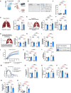
Fig 1
Fig 1
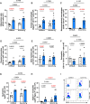
Bacterial clearance is impaired in SCFM2-Scnn1b-Tg mice and increases airway resistance during chronic infection. (A) WT C57BL/6 or Scnn1b-Tg mice were intratracheally inoculated with sterile or 1 × 10⁶ CFU PAO1-laden SCFM2-agar beads. (B) Representative microscopic images of sterile and PAO1-laden SCFM2-agar beads. (C) Bacterial load in mouse lungs 7 days post-infection. Lung bacterial load was determined by CFU/mL. (D–R) Lung function measurements were obtained using the flexiVent (SCIREQ). (D) Inspiratory capacity using a deep inflation technique. (E and F) System resistance (E) and elastance (F) parameters were acquired by the single-frequency forced oscillation maneuver. (G–J) Airway resistance (G), tissue resistance (damping) (H), elastance (I), and hysteresivity (J) were obtained from the low-frequency forced oscillation technique. (J) Representative graph of pressure-volume loop. (L) Static compliance (K) and hysteresis (L) were obtained by a pressure-volume loop. Increased static compliance and hysteresis reflect emphysema and alveolar damage in uninfected Scnn1b-Tg mice compared to WT mice. (M) Representative graph of the forced expiratory volume perturbation. Spirometry measurements from flow-volume loop: FVC (N), FEV0.1 (O), FEV0.1/FVC (P), PEF (Q), and FEF0.1 (R). (C–R) n = 4–5 mice/group, P values indicate two-way ANOVA analyses, red P values indicate P < 0.05, white bars indicate sterile SCFM2 agar bead controls, and blue bars indicate SCFM2 agar beads with PAO1.
Fig 2
Fig 2
Flow cytometry strategy to study the immune response to chronic P. aeruginosa infection in WT SCFM2-C57BL/6 or SCFM2-Scnn1b-Tg mice. (A) WT C57BL/6 or Scnn1b-Tg mice were intratracheally inoculated with sterile or 1 × 10⁶ CFU PAO1-laden SCFM2-agar beads. (B) Gating strategy was used to identify immune cell response at day 7 post infection. Cells were isolated from enzymatically digested mouse lungs, and, after the exclusion of doublets and debris, live and immune cells were identified by LIVE/DEAD staining and CD45 staining. Neutrophils (Ly6G+) were isolated and gated for the Siglec F marker. Then, Ly6G and Siglec F+ cells were selected to differentiate alveolar macrophages (Siglec-F+ and CD11c+) and eosinophils (CD11b+, CD11c, and Siglec-F+). T cells (TCRβ+) were then separated from the rest of Sigle F cells. CD4+ and CD8+ were separated from the double-negative (DN) subset. CD4+ and Foxp3+ cells were isolated, while Foxp3 cells were separated by the CD44 and CD62L markers to identify naïve CD4+ T cells (TCRβ+, CD4+, CD44, and CD62L+), effector CD4+ T cells (TCRβ+, CD4+, CD44+, and CD62L), and central memory CD4+ T cells (TCRβ+, CD4+, CD44+, and CD62L+). CD8+ T cells were also separated with the same markers CD44 and CD62L. Finally, TCR cells were further separated using Ly6C and CD11b markers to identify monocyte-derived macrophages (CD11bHigh, Ly6C+/−, and CD64+), classical monocytes (CD11b+ and Ly6C+), and other myeloid-derived cells (CD11b+ and Ly6C).
Fig 3
Fig 3
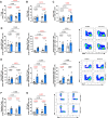
Infection of SCFM2-Scnn1b-Tg mice leads to an increase in atypical neutrophils. (A) Inflammatory cells were increased in both Scnn1b-Tg mice and their WT littermates. (B–E) Different innate cells were upregulated in both genotypes during chronic infection. (B) Alveolar macrophages. (C) Monocyte-derived macrophages. (D) Classical monocytes. (E) Other myeloid cells. (F) Eosinophils were not upregulated during chronic infection with P. aeruginosa. (G) Neutrophils were upregulated during chronic infection but not modulated by the genotype. (H and I) An atypical Siglec F+ neutrophil subset was upregulated in Scnn1b-Tg mice during chronic infection. For (A–H), white bars indicate sterile SCFM2 agar bead controls, blue bars indicate SCFM2 agar beads with PAO1, n = 6 mice/group, mean ± SEM, two-way ANOVA, P < 0.05 are highlighted in red.
Fig 4
Fig 4
Effector T cells increase during P. aeruginosa infection in SCFM2-Scnn1b-Tg-mice. (A) Total T cells were significantly increased during chronic infection and even more in the Scnn1b-Tg mice. (B and C) This increase in T cells was explained by higher numbers of CD4+ (B) and CD8+ (C) T cells. (D) Activation state of CD4+ T cells. No difference was seen in naïve CD4+ T cells. During chronic infection, a significant upregulation of effector T cells was observed in both genotypes, and this increase was greater in Scnn1b-Tg mice compared to their WT littermates. A modest but non-significant increase was detected for central memory T cells in infected mice. (E) Activation state of CD8+ T cells. During chronic infection, a significant increase in naïve CD8+ T cells was observed in Scnn1b-Tg mice. Effector T cells were also increased in both genotypes. A modest increase of central memory CD8 was detected for both genotypes. (F) Regulatory T cells were also increased in all infected mice but not modulated by the genotype. (G) Double-negative (DN) cells were significantly increased in all infected mice and were significantly higher in Scnn1b-Tg mice compared to their WT littermates. For (A–G), white bars indicate sterile SCFM2 agar bead controls, blue bars indicate SCFM2 agar beads with PAO1, n = 6 mice/group, mean ± SEM, two-way ANOVA, P < 0.05 are highlighted in red.
Fig 5
Fig 5
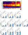
SCFM2-Scnn1b-Tg mice develop exacerbated innate inflammation during chronic infection. (A) Quantification (pg/μg of proteins) of pro- and anti-inflammatory cytokines and chemokines in whole lung lysates of chronically infected mice. (B) Inflammatory cytokines IL-6, IL-1β, and TNF-α were upregulated in all infected mice. IL-1β and TNF-α levels were higher in Scnn1b-Tg mice compared to their WT littermates. (C) Monocytes/macrophages chemoattractant MIP-1α and CXCL10 were significantly upregulated in infected Scnn1b-Tg mice. (D) Neutrophil chemoattractants were significantly upregulated in infected Scnn1b-Tg mice compared to their WT littermates. (E) Lymphocyte chemoattractant MIP-3α was upregulated in all infected mice but was higher in Scnn1b-Tg mice. IL-15 was increased in infected WT C57BL/6 mice only, while IL-16 was only upregulated in Scnn1b-Tg mice. For (B–E), white bars indicate sterile SCFM2 agar bead controls, blue bars indicate SCFM2 agar beads with PAO1, n = 4–5 mice/group, *P < 0.05, **P < 0.01, and ***P < 0.001. See Table S1 for statistical tests used and exact P values.
Fig 6
Fig 6
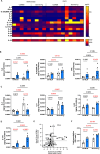
Chronic infection leads to dysfunctional lymphoid-mediated inflammation in SCFM2-Scnn1b-Tg mice. (A) Quantification (pg/μg of proteins) of types 1, 2, and 3 inflammation cytokines and chemokines in whole lung lysates of chronically infected mice. (B) Type 1 inflammation lymphokines IL-2, IL-27p/28/IL-30, and IFN-γ were significantly upregulated in infected Scnn1b-Tg mice. (C) Type 2 inflammation lymphokines IL-4 and IL-5 were upregulated in uninfected Scnn1b-Tg mice. Although IL-4 was downregulated during infection, IL-5 levels were maintained. IL-33 was upregulated in Scnn1b-Tg mice during chronic infection. (D) Type 3 inflammation cytokine IL-17 was upregulated in all mice and further increased in Scnn1b-Tg mice. (E) Z-scores highlight type 2 and lymphoid inflammation in uninfected Scnn1b-Tg mice compared to their WT littermates. IL-17 is the most differentially upregulated cytokine in these mice during infection. (F) Scnn1b-Tg have higher lung tissue damage at baseline. Chronic infection caused increased lipid peroxidation in all infected mice but was greater in Scnn1b-Tg mice. For (B–D and F), white bars indicate sterile SCFM2 agar bead controls, blue bars indicate SCFM2 agar beads with PAO1, n = 4–5 mice/group, *P < 0.05, **P < 0.01, ***P < 0.001, ****P < 0.0001. See Table S1 for statistical tests used and exact P values.
Fig 7
Fig 7
Summary of lung inflammation in SCFM2-C57BL/6 and SCFM2-Scnn1b-Tg mice during chronic infection with P. aeruginosa. Healthy lungs from C57BL/6 mice have surveilling alveolar macrophages. Uninfected Scnn1b-Tg mice show underlying inflammation characterized by the presence of alveolar and monocyte-derived macrophages, monocytes, other myeloid cells, and effector T cells. Conventional and Siglec F+ neutrophils are also present in the BAL of uninfected Scnn1b-Tg mice. Type 2 inflammation, demonstrated by the presence of eosinophils and IL-4 and IL-5, is present at baseline in the Scnn1b-Tg lung environment. During chronic infection, both SCFM2-C57BL/6 and SCFM2-Scnn1b-Tg immune responses are characterized by infiltration of innate and T cells and high levels of types 1 and 3 inflammation cytokines and chemokines (IL-1β, IL-2, IL-6, IL-17, TNFα, IFNγ, MIP-1α, MIP-2, MIP-3α, and KC/GRO). In addition to being exacerbated, SCFM2-Scnn1b-Tg inflammation is characterized by a sustained type 2 inflammation, a marked IL-17/neutrophil interplay, and the recruitment of unconventional Siglec F+ neutrophils. Higher inflammation is associated with higher lung tissue damage in SCFM2-Scnn1b-Tg mice. Cytokines in red font are cytokines expressed in the specific genotypes. Red arrows indicate where the cytokine production is increased relative to the other genotype.

Update of

References

    1. Garcia-Clemente M, de la Rosa D, Máiz L, Girón R, Blanco M, Olveira C, Canton R, Martinez-García MA. 2020. Impact of Pseudomonas aeruginosa infection on patients with chronic inflammatory airway diseases. J Clin Med 9:3800. doi: 10.3390/jcm9123800 - DOI - PMC - PubMed
    1. Stanford GE, Dave K, Simmonds NJ. 2021. Pulmonary exacerbations in adults with cystic fibrosis. Chest 159:93–102. doi: 10.1016/j.chest.2020.09.084 - DOI - PMC - PubMed
    1. Sanders DB, Bittner RCL, Rosenfeld M, Redding GJ, Goss CH. 2011. Pulmonary exacerbations are associated with subsequent FEV1 decline in both adults and children with cystic fibrosis. Pediatr Pulmonol 46:393–400. doi: 10.1002/ppul.21374 - DOI - PubMed
    1. Rossi E, Falcone M, Molin S, Johansen HK. 2018. High-resolution in situ transcriptomics of Pseudomonas aeruginosa unveils genotype independent patho-phenotypes in cystic fibrosis lungs. Nat Commun 9:3459. doi: 10.1038/s41467-018-05944-5 - DOI - PMC - PubMed
    1. Vaillancourt M, Galdino ACM, Limsuwannarot SP, Celedonio D, Dimitrova E, Broerman M, Bresee C, Doi Y, Lee JS, Parks WC, Jorth P. 2023. A compensatory RNase E variation increases iron piracy and virulence in multidrug-resistant Pseudomonas aeruginosa during macrophage infection. PLoS Pathog 19:e1010942. doi: 10.1371/journal.ppat.1010942 - DOI - PMC - PubMed

Publication types

MeSH terms

Substances