. 2025 Aug;120(4):761-777.
doi: 10.1007/s00395-025-01119-8. Epub 2025 Jun 13.

Direction-dependent contributions of cardiac myofilament networks to myocardial passive stiffness reveal a major disparity for titin

Affiliations

Direction-dependent contributions of cardiac myofilament networks to myocardial passive stiffness reveal a major disparity for titin

Felix A Wagner et al. Basic Res Cardiol. 2025 Aug.

Abstract

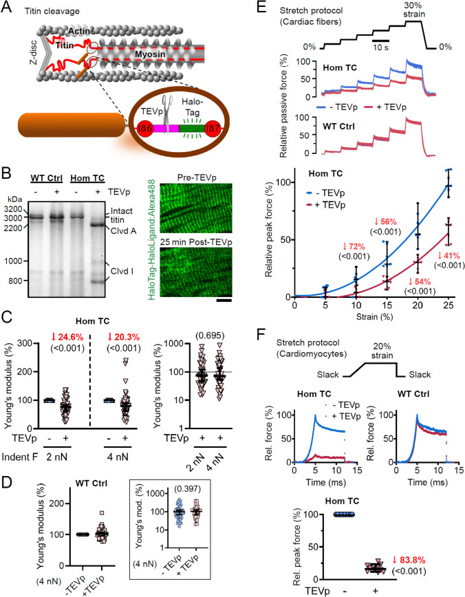
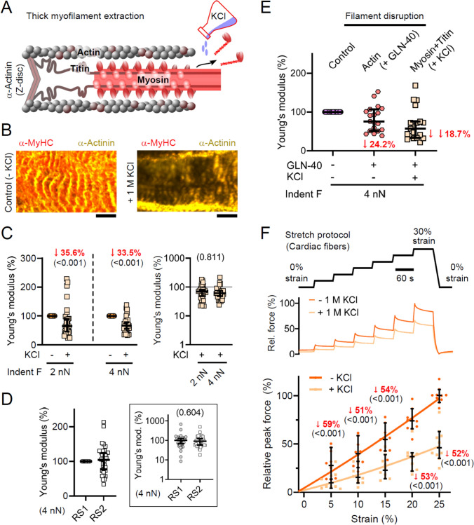
Progressive myocardial dysfunction in patients with heart failure often involves alterations in myocardial passive stiffness, yet the underlying mechanisms remain incompletely understood. While passive stiffness in the longitudinal direction has been extensively characterized via uniaxial tensile stretching of cardiac specimens, transverse stiffness has received far less attention despite its equal mechanical importance. In this study, we combined atomic force microscopy nanoindentation with stretching assays on myocardial preparations to quantify the relative contributions of the three myofilament networks - actin, myosin, and titin - to passive stiffness in both transverse and longitudinal orientations. We employed a transgenic mouse model in which titin's elastic springs contain a tobacco etch virus protease (TEVp) recognition site, enabling selective and acute titin cleavage upon TEVp treatment. Actin filaments were severed using a calcium-independent gelsolin fragment, and myosin filaments were dissociated by high-salt extraction. Along the longitudinal axis, titin accounted for over 50% of total passive stiffness in both cardiac fiber bundles and isolated cardiomyocytes across most physiological strain ranges, whereas actin contributed under 35% overall - and only 15-20% within the collagen-containing fiber bundles. In contrast, in the transverse axis, titin and actin each contributed approximately 20-26% of passive stiffness in cardiac slices under varying compression forces. The myosin-titin composite thick-filament network contributed ~ 55% longitudinally but only ~ 35% transversely. These results reveal pronounced, direction-dependent differences in myofilament contributions to myocardial passive stiffness, with titin exhibiting the greatest disparity. Our findings deepen our understanding of the myocardium's multidimensional mechanics and may inform therapeutic strategies to ameliorate pathological cardiac stiffening.

Keywords: Actin; Atomic force microscopy; Myocardium; Myosin; Passive stiffness; Titin.


Conflict of interest statement

Declarations. Conflict of interest: All authors declare that they have no conflict of interest related to this manuscript. Ethics: We obtained permission for all animal procedures from the local animal welfare authority (Landesamt für Natur, Umwelt und Verbraucherschutz Nordrhein-Westfalen, LANUV NRW, 81–02.04.2019.A472). The manuscript does not contain clinical studies or patient data.

Figures

Fig. 1
Fig. 1
Measuring transverse passive stiffness in myocardial tissue slices with atomic force microscopy (AFM). A Schematic of the AFM setup (left), with blue dots marking measurement points in the red-lined piezo range, and representative force-height curve (right), with Young’s modulus calculated from the 2.00–2.25 nN and 4.00–4.25 nN force ranges (purple). B Representative fluorescence image of cardiac tissue section from a homozygous (Hom) titin-cleavage (TC) mouse labeled for titin with HaloLigand:AlexaFluor-488; the AFM cantilever (here, for demonstration purposes, Arrow-TL1, Nanoworld) and indenter position (white arrow) are indicated. Scale bar, 20 µm. C Histogram summarizing local Young’s moduli recorded in wRS (n = 280 locations, N = 33 slices) at 4.00–4.25 nN, with an inset heat map of repeated measurements (Repeats 1–3) of the same region and mean values; scale bars, 10 µm
Fig. 2
Fig. 2
Contribution of actin filaments to transverse and longitudinal passive stiffness. A Schematic of GLN-40-mediated actin severing. B Permeabilized mouse LV cardiac samples treated for 60 min in wRS ± 50 µg GLN-40 and imaged by IF microscopy (left) using antibodies to actin (phalloidin-Alexa-647, magenta) and α-actinin (EA53 + AlexaFluor-488, yellow); scale bars, 5 µm. SDS-PAGE (right): homogenized TC fibers incubated for 2 h in iRS ± 25 µg GLN-40, pellet and supernatant separated, Coomassie-stained. C AFM nanoindentation of permeabilized WT cardiac slices (n = 78 measurements, N = 4 slices) before/after 60 min wRS + 50 µg GLN-40. Left: post/pre Young’s modulus at 2.00–2.25 nN and 4.00–4.25 nN indentation force; right: Young’s modulus (logarithmic scale) for GLN-40-treated samples, normalized to median pre-treatment. Data are median ± 95% CI. LME ANOVA with treatment and force level as fixed factors, measurement location as random factor. D AFM of WT slices (n = 30 measurements, N = 3 slices) before/after 60 min wRS alone (RS1 vs. RS2; these represent the same wRS but at different time points), recorded at 4.00–4.25 nN indentation force. Inset: Young’s modulus (logarithmic scale) relative to median RS1 condition. Data are median ± 95% CI. LME ANOVA with treatment as fixed factor, location random. E Passive force-extension on permeabilized TC cardiac fiber bundles (n = 12 measurements, N = 6 mice) before/after 30 min iRS + 50 µg GLN-40. Top: stretch protocol; middle: representative traces; bottom: peak force (median ± 95% CI) normalized to pre-treatment maximum, with second-order polynomial fits. LME ANOVA with treatment and strain (4–20%) as fixed factors, fiber as random factor; significant median changes in red (p in parentheses). F Passive force of permeabilized isolated cardiomyocytes before/after 30 min iRS + 25 µg GLN-40. Top: stretch protocol; middle left: phase image with ROI used for sarcomere length measurements (scale bars, 20 µm); middle right: force traces; bottom: average peak force (median ± 95% CI; n = 12 cardiomyocytes, N = 5 mice) normalized to pre-treatment. One-sample t-test; significant median change in red (p in parenthesis)
Fig. 3
Fig. 3
Contribution of titin springs to transverse and longitudinal passive stiffness. A Schematic of a Hom TC half-sarcomere illustrating the TEVp recognition site-HaloTag cassette in titin’s elastic I-band (between immunoglobulin domains I86 and I87); scissors denote TEVp cleavage. B Left: SDS-PAGE of permeabilized WT control (Ctrl) and Hom TC cardiac tissue after incubation in iRS + TEVp (100 mM DTT) or iRS (+ DTT) alone. Right: Hom TC cardiac section before/after TEVp, HaloTag labeled with HaloLigand-AlexaFluor-488; scale bar, 10 µm. C AFM nanoindentation of permeabilized Hom TC slices before/after 25 min wRS + TEVp (n = 94 measurements, N = 7 slices). Left: post/pre Young’s modulus at 2.00–2.25 nN and 4.00–4.25 nN indentation force; right: Young’s modulus (logarithmic scale) for TEVp-treated samples normalized to median pre-treatment. Data are median ± 95% CI. Analysis by LME ANOVA (treatment and force level fixed; location random). D AFM of WT slices (n = 45 measurements, N = 5 slices) before/after identical treatment, recorded at 4.00–4.25 nN indentation force. Inset: Young’s modulus (logarithmic scale) relative to median—TEVp condition. Data are median ± 95% CI. LME ANOVA with treatment as fixed factor, location random. E Passive force-extension on permeabilized Hom TC cardiac fiber bundles (n = 7 measurements, N = 5 mice) before/after 10 min iRS + TEVp, with WT fibers as controls. Top: stretch protocol; middle: representative traces (for WT, the—TEVp and + TEVp traces are superimposed); bottom: peak force of Hom TC fibers before/after treatment (median ± 95% CI) normalized to pre-treatment at 25% strain. LME ANOVA (treatment and strain fixed; fiber random); fits are second-order polynomials; significant median changes in red (p in parentheses). F Passive force of permeabilized isolated cardiomyocytes before/after 10 min iRS + TEVp. Top: stretch protocol; middle: representative traces for a Hom TC (left) and WT (right); bottom: peak force (median ± 95% CI; n = 11 cells, N = 5 mice) normalized to pre-treatment in Hom TC cells. One-sample t-test vs. 100; significant median change in red (p in parenthesis)
Fig. 4
Fig. 4
Contribution of titin-myosin composite (thick) filaments to transverse and longitudinal passive stiffness. A Schematic of KCl-induced thick-filament depolymerization. B IF images of WT cardiac tissue after 10 min in wRS alone (left) or wRS + 1 M KCl (right). Myosin (anti-α-MHC, Cy3) is shown in orange; α-actinin (EA 53, AlexaFluor-488) in yellow. Scale bars, 5 µm. C AFM nanoindentation of permeabilized WT slices (n = 63 measurements, N = 5 slices) before/after 10 min wRS + 1 M KCl. Left: post-/pre-treatment Young’s modulus at 2.00–2.25 nN and 4.00–4.25 nN indentation force; right: post-treatment Young’s moduli (logarithmic scale) relative to the median pre-treatment value. Data are median ± 95% CI. LME ANOVA with treatment and compression as fixed factors; location as random factor. D AFM of WT slices (n = 35 measurements, N = 5 slices) before/after 10 min wRS alone (RS1 vs. RS2), recorded at 4.00–4.25 nN indentation force. Inset: Young’s modulus (logarithmic scale) relative to median RS1 condition. Data are median ± 95% CI. LME ANOVA with treatment as fixed factor, location random. E AFM nanoindentation of WT slices (n = 20 measurements, N = 2 slices) at 4.00–4.25 nN indentation force: control (wRS), after 60 min wRS + 50 µg GLN-40, and after an additional 10 min wRS + 1 M KCl; Youngs’ moduli normalized to control condition. Data are median ± 95% CI. F Passive force-extension of permeabilized WT fiber bundles (n = 10 measurements, N = 6 mice) before/after 60 min wRS + 1 M KCl. Top: stretch protocol; middle: representative traces; bottom: peak force before/after treatment (median ± 95% CI) normalized to pre-treatment at 25% strain. LME ANOVA (treatment and strain fixed; fiber random); fits are second-order polynomials; significant median changes in red (p in parentheses)
Fig. 5
Fig. 5
Directional contributions to passive stiffness from actin, titin, and myosin-titin composite filaments. A Relative stiffness of individual filament systems measured under high-strain (20%) conditions in isolated cardiomyocytes, in cardiac fiber bundles at both high (20%) and low (10–12%) strain, and by AFM nanoindentation of cardiac tissue slices subjected to high (4.00–4.25 nN) and low (2.00–2.25 nN) compressive loads. B Longitudinal-to-transverse stiffness quotient (LTQ) computed from the data in (A) for each myofilament type, comparing measurements in fiber bundles versus cardiac slices. Left: LTQ at low strain (10–12%) or low compression (2.00–2.25 nN) for actin, titin, and myosin-titin composite filaments; right: LTQ at high strain (20%) or high compression (4.00–4.25 nN) for the same three components

References

    1. Akiyama N, Ohnuki Y, Kunioka Y, Saeki Y, Yamada T (2006) Transverse stiffness of myofibrils of skeletal and cardiac muscles studied by atomic force microscopy. J Physiol Sci 56:145–151. 10.2170/physiolsci.RP003205 - PubMed
    1. Anderson BR, Granzier HL (2012) Titin-based tension in the cardiac sarcomere: molecular origin and physiological adaptations. Prog Biophys Mol Biol 110:204–217. 10.1016/j.pbiomolbio.2012.08.003 - PMC - PubMed
    1. Armstrong PW, Lam CSP, Anstrom KJ, Ezekowitz J, Hernandez AF, O’Connor CM, Pieske B, Ponikowski P, Shah SJ, Solomon SD, Voors AA, She L, Vlajnic V, Carvalho F, Bamber L, Blaustein RO, Roessig L, Butler J (2020) Effect of vericiguat vs placebo on quality of life in patients with heart failure and preserved ejection fraction: the VITALITY-HFpEF randomized clinical trial. JAMA 324:1512–1521. 10.1001/jama.2020.15922 - PMC - PubMed
    1. Azeloglu EU, Costa KD (2010) Cross-bridge cycling gives rise to spatiotemporal heterogeneity of dynamic subcellular mechanics in cardiac myocytes probed with atomic force microscopy. Am J Physiol Heart Circ Physiol 298:H853–H860. 10.1152/ajpheart.00427.2009 - PubMed
    1. Bishu K, Hamdani N, Mohammed SF, Kruger M, Ohtani T, Ogut O, Brozovich FV, Burnett JC Jr, Linke WA, Redfield MM (2011) Sildenafil and B-type natriuretic peptide acutely phosphorylate titin and improve diastolic distensibility in vivo. Circulation 124:2882–2891. 10.1161/CIRCULATIONAHA.111.048520 - PMC - PubMed

LinkOut - more resources